Copyright
©2010 Baishideng Publishing Group Co.
World J Radiol. Oct 28, 2010; 2(10): 399-404
Published online Oct 28, 2010. doi: 10.4329/wjr.v2.i10.399
Published online Oct 28, 2010. doi: 10.4329/wjr.v2.i10.399
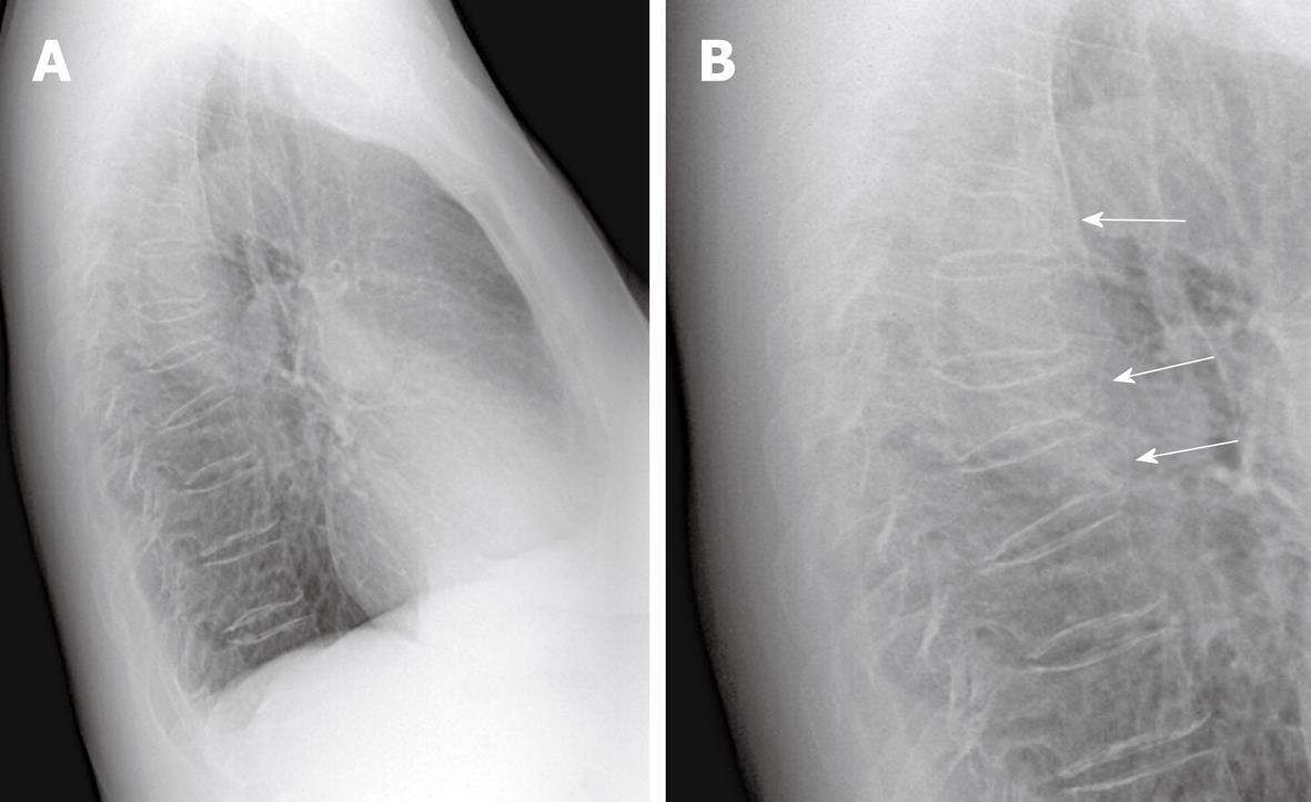
Figure 1 Incidental vertebral compression fractures on chest radiograph.
A: Lateral radiograph of the chest of a 65 year old woman studied for persistent cough. No relevant pulmonary abnormality and mild cardiomegaly were noted in the frontal radiograph (not shown); B: Close up on the vertebral column shows the presence of a mild grade biconcave fracture of T6, severe grade wedge fracture of T8 and moderate grade fracture of T9 (arrows).
- Citation: Bartalena T, Rinaldi MF, Modolon C, Braccaioli L, Sverzellati N, Rossi G, Rimondi E, Busacca M, Albisinni U, Resnick D. Incidental vertebral compression fractures in imaging studies: Lessons not learned by radiologists. World J Radiol 2010; 2(10): 399-404
- URL: https://www.wjgnet.com/1949-8470/full/v2/i10/399.htm
- DOI: https://dx.doi.org/10.4329/wjr.v2.i10.399


