Copyright
©2010 Baishideng Publishing Group Co.
World J Radiol. Oct 28, 2010; 2(10): 384-398
Published online Oct 28, 2010. doi: 10.4329/wjr.v2.i10.384
Published online Oct 28, 2010. doi: 10.4329/wjr.v2.i10.384
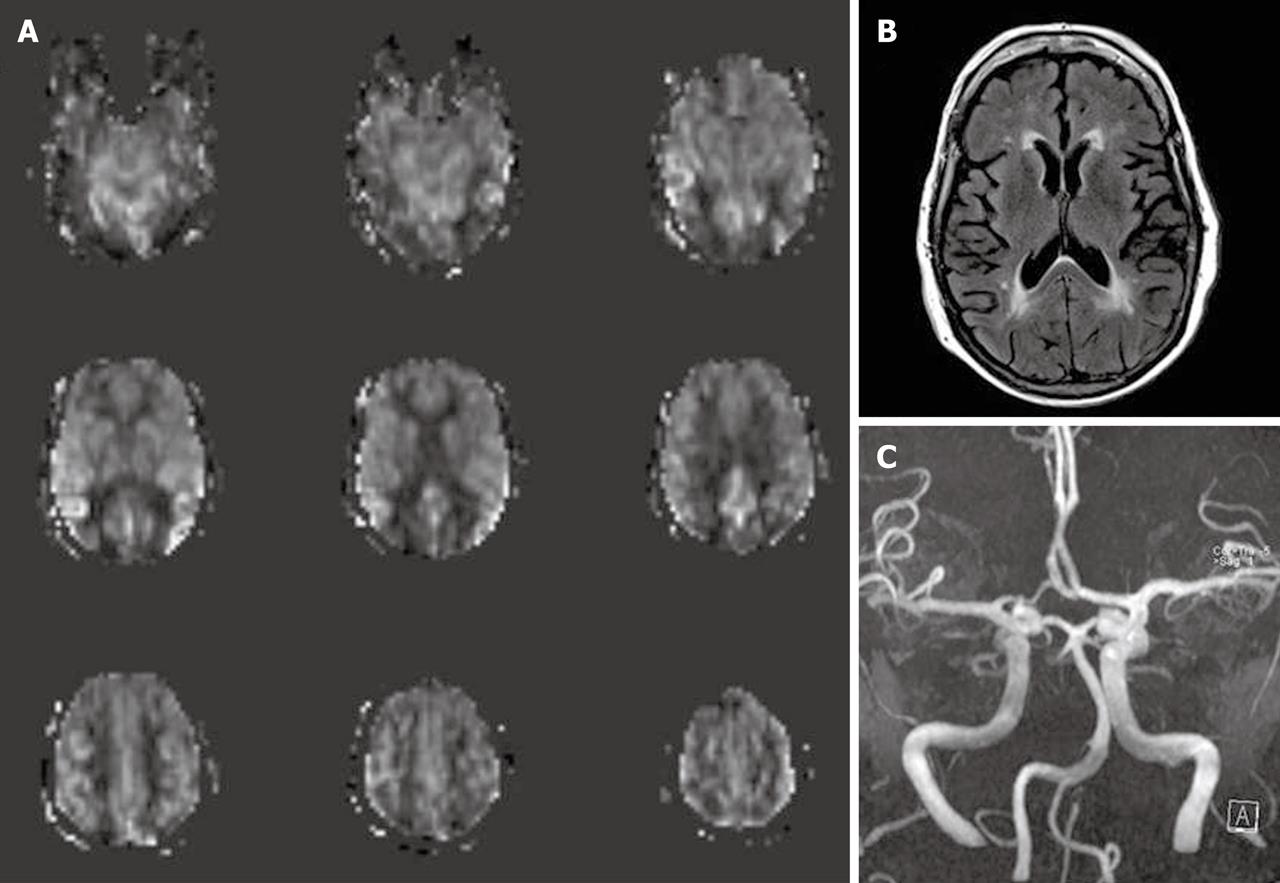
Figure 6 A 77-year-old female underwent magnetic resonance imaging of the brain due to cognitive impairment.
Arterial spin labeling cerebral blood flow maps (A) show a borderzone sign, seen as low signal intensity in arterial watershed regions with serpiginous high signal intensity in the surrounding cortex. FLAIR images (B) reveal nonspecific high signal intensity in periventricular deep white matter without other abnormality. Magnetic resonance angiography (MRA) of the brain (C), MRA of the neck and dynamic susceptibility contrast magnetic resonance perfusion (not shown) were unremarkable.
- Citation: Petcharunpaisan S, Ramalho J, Castillo M. Arterial spin labeling in neuroimaging. World J Radiol 2010; 2(10): 384-398
- URL: https://www.wjgnet.com/1949-8470/full/v2/i10/384.htm
- DOI: https://dx.doi.org/10.4329/wjr.v2.i10.384


