Copyright
©2010 Baishideng Publishing Group Co.
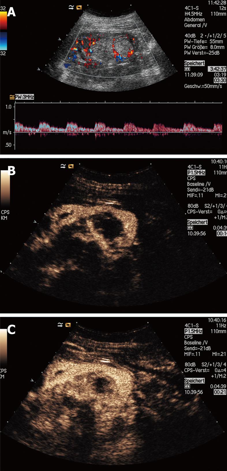
Figure 2 Histologically proven angiomyolipoma with typical central artery (which has been also described in some oncocytoma).
Doppler US analysis reveals a relatively low resistance index (A); In CEUS the lesion shows a hypovascular enhancement (B, C).
- Citation: Ignee A, Straub B, Schuessler G, Dietrich CF. Contrast enhanced ultrasound of renal masses. World J Radiol 2010; 2(1): 15-31
- URL: https://www.wjgnet.com/1949-8470/full/v2/i1/15.htm
- DOI: https://dx.doi.org/10.4329/wjr.v2.i1.15


