Copyright
©2010 Baishideng Publishing Group Co.
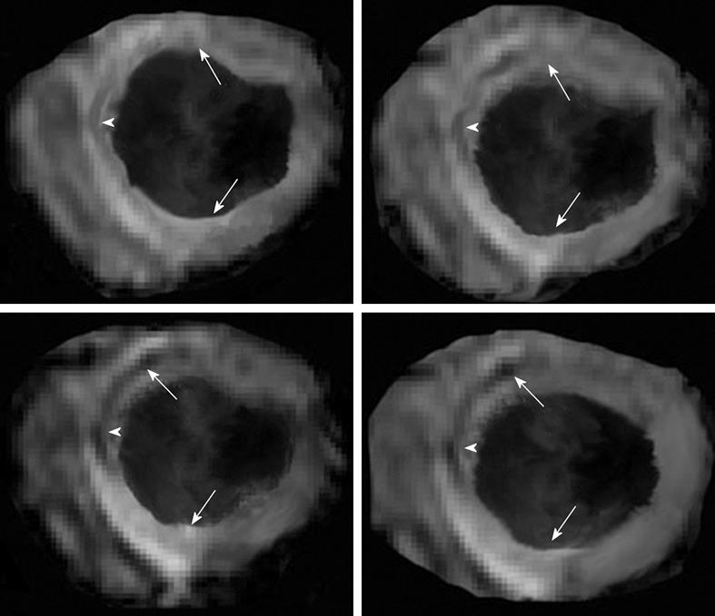
Figure 7 Multislice T2*-weighted (susceptibility) turbo spin echo images show severe interstitial hemorrhage in the core (arrowheads) of the hyperintense edematous area at risk (arrows) 3 d after reperfusion.
- Citation: Saeed M, Hetts S, Wilson M. Reperfusion injury components and manifestations determined by cardiovascular MR and MDCT imaging. World J Radiol 2010; 2(1): 1-14
- URL: https://www.wjgnet.com/1949-8470/full/v2/i1/1.htm
- DOI: https://dx.doi.org/10.4329/wjr.v2.i1.1


