Copyright
©The Author(s) 2025.
World J Radiol. Mar 28, 2025; 17(3): 103822
Published online Mar 28, 2025. doi: 10.4329/wjr.v17.i3.103822
Published online Mar 28, 2025. doi: 10.4329/wjr.v17.i3.103822
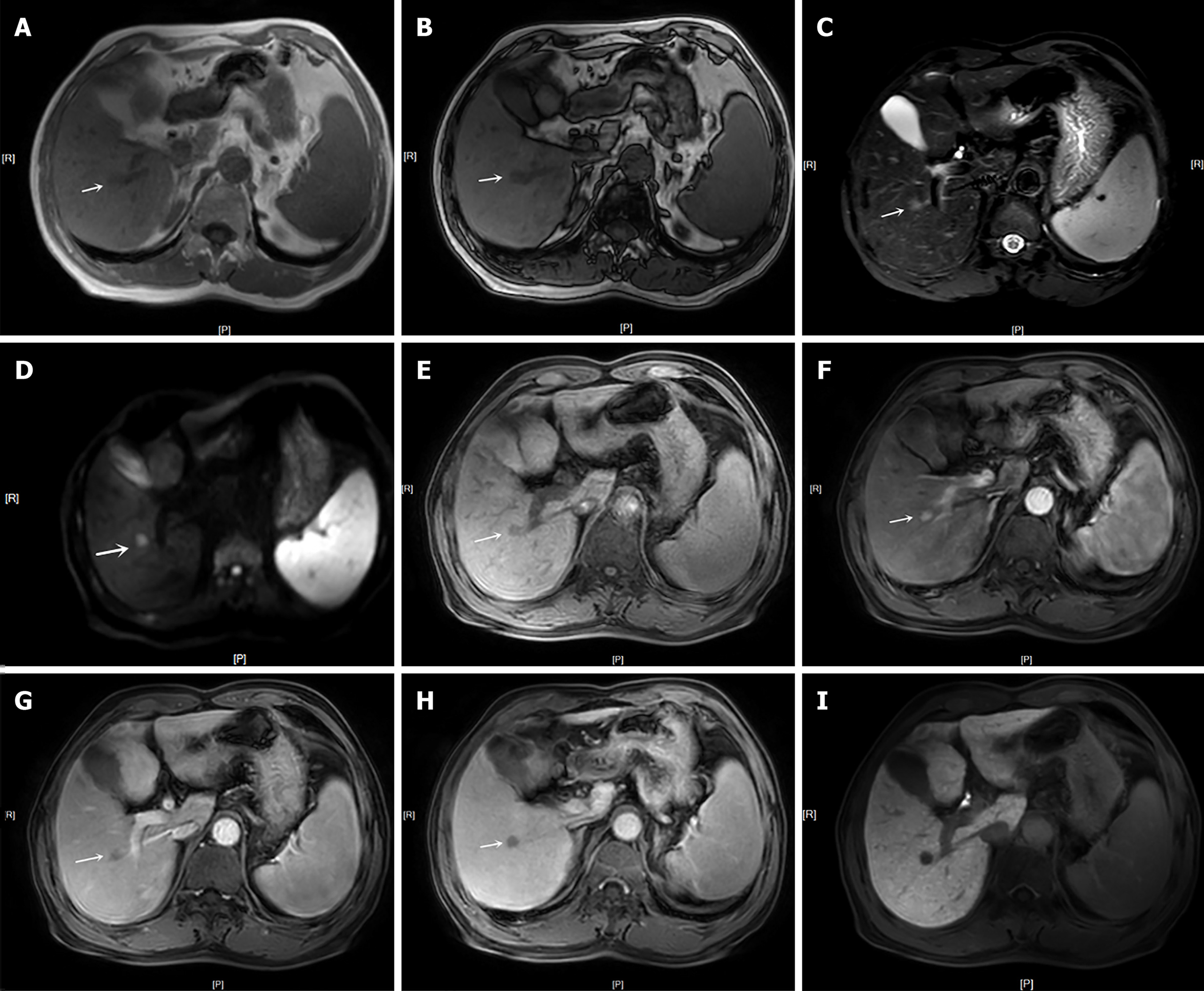
Figure 2 Axial images obtained with gadoxetic acid–enhanced magnetic resonance imaging in a 62-year-old man with cirrhosis by chronic B-viral hepatitis and surgically confirmed hepatocellular carcinoma.
This lesion was assigned as LR-4 and could not be classified as LR-5 using Liver Imaging Reporting and Data System version 2018. In contrast, our modified LR-5 criteria or alternative criteria (LR-5-M1, LR-5-M2, LR-5-M8 and LR-5-M9) by utilizing independently significant ancillary features (nonrim arterial phase hyperenhancement, nonperipheral washout, mild-moderate T2 hyperintensity and transition phase hypointensity) helped achieve the correct diagnosis of hepatocellular carcinoma. A and B: In-/out-of-phase T1-weighted image showed a 0.9 cm-diameter mass in segment VIII of the liver (white arrow); C: T2-weighted image showed mild-moderate T2 hyperintensity in the lesion; D: Diffusion-weighted imaging showed restricted diffusion in the lesion; E: Pre-enhanced phase showed hypointensity in the lesion; F: Late artery phase showed nonrim arterial phase hyperenhancement in the lesion; G: Portal venous phase showed nonperipheral washout in the lesion; H and I: The transition phase and hepatobiliary phase showed hypointensity in the lesion (white arrow).
- Citation: Song Y, Zhang YY, Yu Q, Ma R, Xiao Y, Shen JK, Wei CG. Modified LR-5 criteria based on gadoxetic acid can improve the sensitivity in the diagnosis of hepatocellular carcinoma. World J Radiol 2025; 17(3): 103822
- URL: https://www.wjgnet.com/1949-8470/full/v17/i3/103822.htm
- DOI: https://dx.doi.org/10.4329/wjr.v17.i3.103822


