Copyright
©The Author(s) 2025.
World J Radiol. Feb 28, 2025; 17(2): 104518
Published online Feb 28, 2025. doi: 10.4329/wjr.v17.i2.104518
Published online Feb 28, 2025. doi: 10.4329/wjr.v17.i2.104518
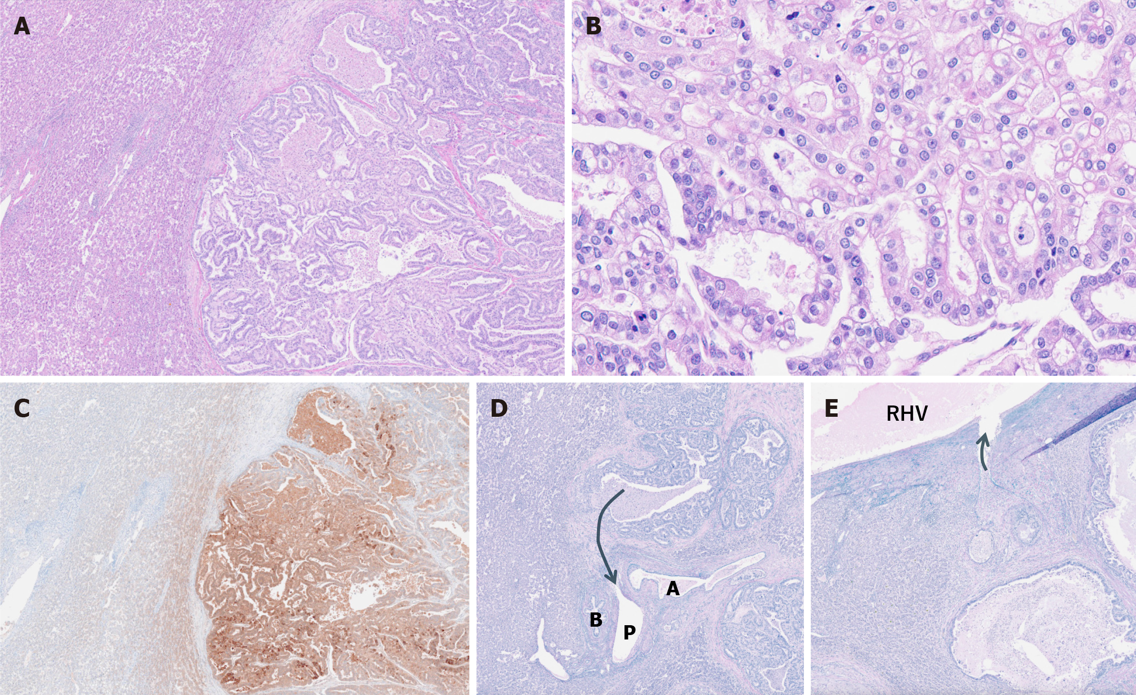
Figure 4 Histopathological images of the surgical specimen.
A: Atypical cells with clear cytoplasm were observed, proliferating in a tubular to papillary pattern (hematoxylin and eosin staining, magnification × 25); B: Higher magnification of the same area as in Figure 4A (hematoxylin and eosin staining, magnification × 200); C: Immunohistochemistry demonstrated that the tumor cells were positive for alpha-fetoprotein; D: Elastica Van Gieson staining revealed invasion of the portal vein (arrow); E: Elastica Van Gieson staining also revealed invasion of the right hepatic vein of the liver parenchyma surrounding the tumor (arrow). RHV: Right hepatic vein.
- Citation: Irizato M, Minamiguchi K, Fujita Y, Yamaura H, Onaya H, Taiji R, Tanaka T, Inaba Y. Distinctive imaging features of liver metastasis from gastric adenocarcinoma with enteroblastic differentiation: A case report. World J Radiol 2025; 17(2): 104518
- URL: https://www.wjgnet.com/1949-8470/full/v17/i2/104518.htm
- DOI: https://dx.doi.org/10.4329/wjr.v17.i2.104518


