Copyright
©The Author(s) 2025.
World J Radiol. Jan 28, 2025; 17(1): 99207
Published online Jan 28, 2025. doi: 10.4329/wjr.v17.i1.99207
Published online Jan 28, 2025. doi: 10.4329/wjr.v17.i1.99207
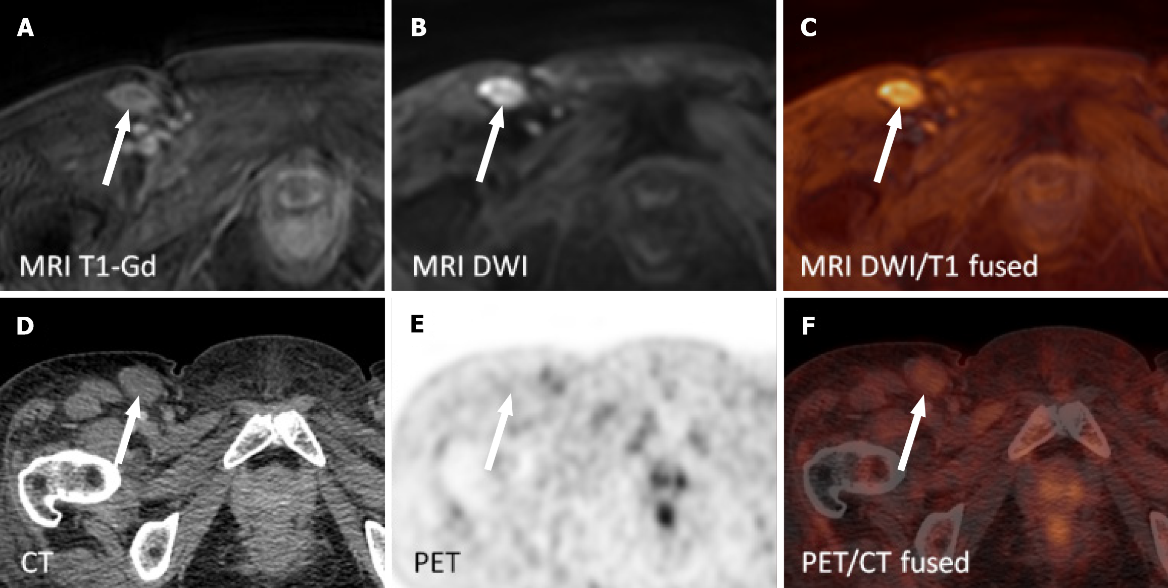
Figure 3 Non-avid right inguinal lymph node on fluorodeoxyglucose positron emission tomography/computed tomography was reported positive on whole-body magnetic resonance imaging five days later.
The patient underwent extirpation of a lymph node from the right groin with persisting reactive changes and seroma. A: Contrast-enhanced T1 weighted single breath-hold images; B: Diffusion weighted images; C: Diffusion weighted images and contrast-enhanced T1 weighted images on magnetic resonance imaging (fused); D: Computed tomography; E: Positron emission tomography; F: Positron emission tomography/computed tomography images (fused). MRI: Magnetic resonance imaging; T1-Gd: Contrast-enhanced T1 weighted single breath-hold images; DWI: Diffusion weighted images; PET/CT: Positron emission tomography/computed tomography.
- Citation: Lambert L, Wagnerova M, Vodicka P, Benesova K, Zogala D, Trneny M, Burgetova A. Whole-body magnetic resonance imaging provides accurate staging of diffuse large B-cell lymphoma, but is less preferred by patients. World J Radiol 2025; 17(1): 99207
- URL: https://www.wjgnet.com/1949-8470/full/v17/i1/99207.htm
- DOI: https://dx.doi.org/10.4329/wjr.v17.i1.99207


