Copyright
©The Author(s) 2025.
World J Radiol. Jan 28, 2025; 17(1): 97255
Published online Jan 28, 2025. doi: 10.4329/wjr.v17.i1.97255
Published online Jan 28, 2025. doi: 10.4329/wjr.v17.i1.97255
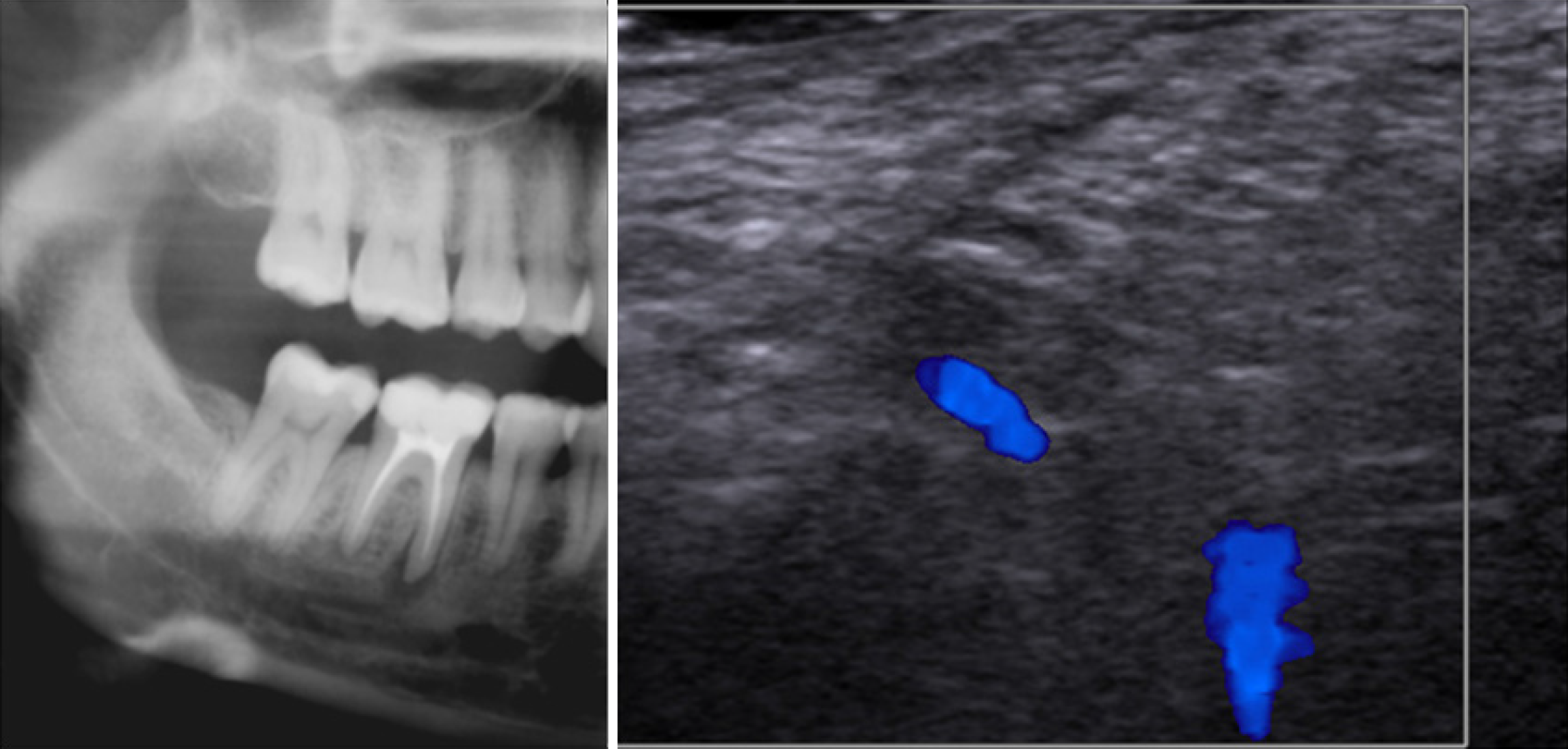
Figure 7 Cropped panoramic radiography (left) and laser doppler ultrasonographic image of a 30-year-old female patient referred for slight percussion sensitivity in the right mandibular first molar region.
On panoramic radiography, a well-defined radiolucent lesion on the mesial root of the first mandibular right molar was observed. Laser Doppler ultrasonographic evaluation revealed that there was blood flow in the region suggestive of a granuloma, which was proved by histopathological examination.
- Citation: Kamburoğlu K. Trends in dentomaxillofacial radiology. World J Radiol 2025; 17(1): 97255
- URL: https://www.wjgnet.com/1949-8470/full/v17/i1/97255.htm
- DOI: https://dx.doi.org/10.4329/wjr.v17.i1.97255


