Copyright
©The Author(s) 2025.
World J Radiol. Jan 28, 2025; 17(1): 97255
Published online Jan 28, 2025. doi: 10.4329/wjr.v17.i1.97255
Published online Jan 28, 2025. doi: 10.4329/wjr.v17.i1.97255
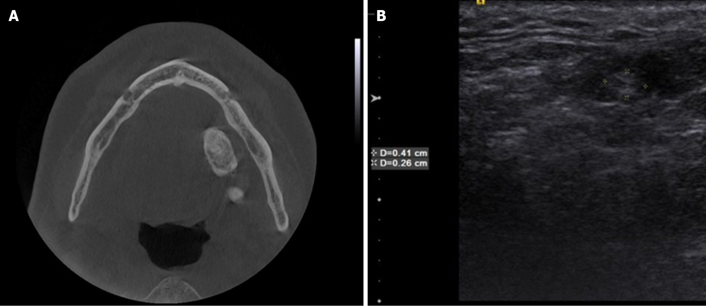
Figure 6 Axial cone beam CT and ultrasonographic measurement images.
A and B: Axial cone beam CT (A) and ultrasonographic measurement (B) images of a 25-year-old female patient referred to our clinic with the complaints of pain and swelling of the cheek after meals. The radiopaque lesion on cone beam CT and hyperechoic lesion on ultrasound images in the submandibular region were suggestive of a submandibular sialolithiasis.
- Citation: Kamburoğlu K. Trends in dentomaxillofacial radiology. World J Radiol 2025; 17(1): 97255
- URL: https://www.wjgnet.com/1949-8470/full/v17/i1/97255.htm
- DOI: https://dx.doi.org/10.4329/wjr.v17.i1.97255


