Copyright
©The Author(s) 2025.
World J Radiol. Jan 28, 2025; 17(1): 97255
Published online Jan 28, 2025. doi: 10.4329/wjr.v17.i1.97255
Published online Jan 28, 2025. doi: 10.4329/wjr.v17.i1.97255
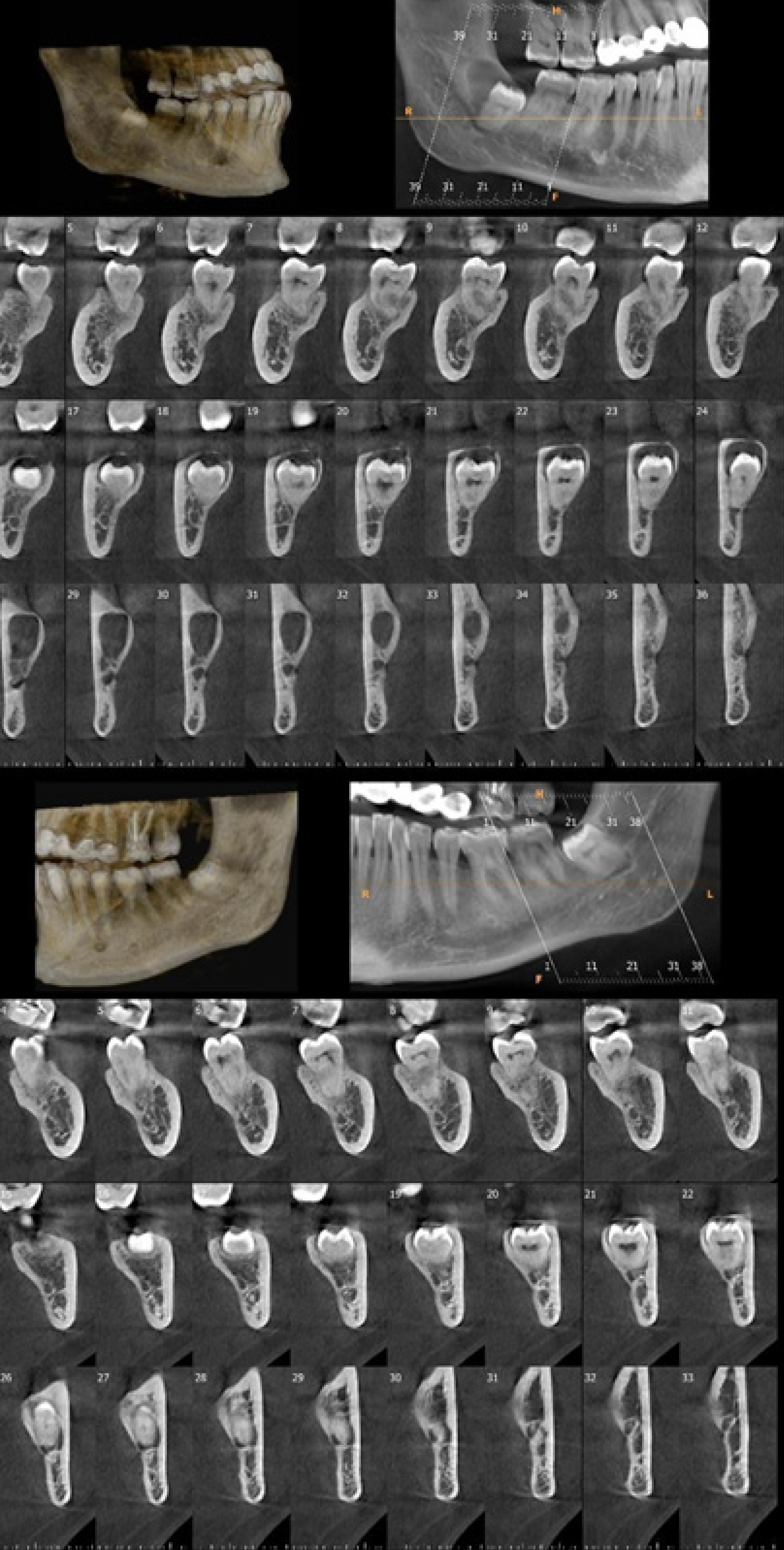
Figure 5 Cone beam CT images of mandibular right (upper image) and left (lower image) third molar teeth of a 25-year-old female patient referred for persistent pain on both retromolar sides.
Three-dimensional bone model, panoramic, and cross-sectional images showed a dentigerous cyst related to the mandibular right third molar tooth and direct contact of mandibular left third molar tooth root with mandibular canal.
- Citation: Kamburoğlu K. Trends in dentomaxillofacial radiology. World J Radiol 2025; 17(1): 97255
- URL: https://www.wjgnet.com/1949-8470/full/v17/i1/97255.htm
- DOI: https://dx.doi.org/10.4329/wjr.v17.i1.97255


