Copyright
©The Author(s) 2025.
World J Radiol. Jan 28, 2025; 17(1): 97255
Published online Jan 28, 2025. doi: 10.4329/wjr.v17.i1.97255
Published online Jan 28, 2025. doi: 10.4329/wjr.v17.i1.97255
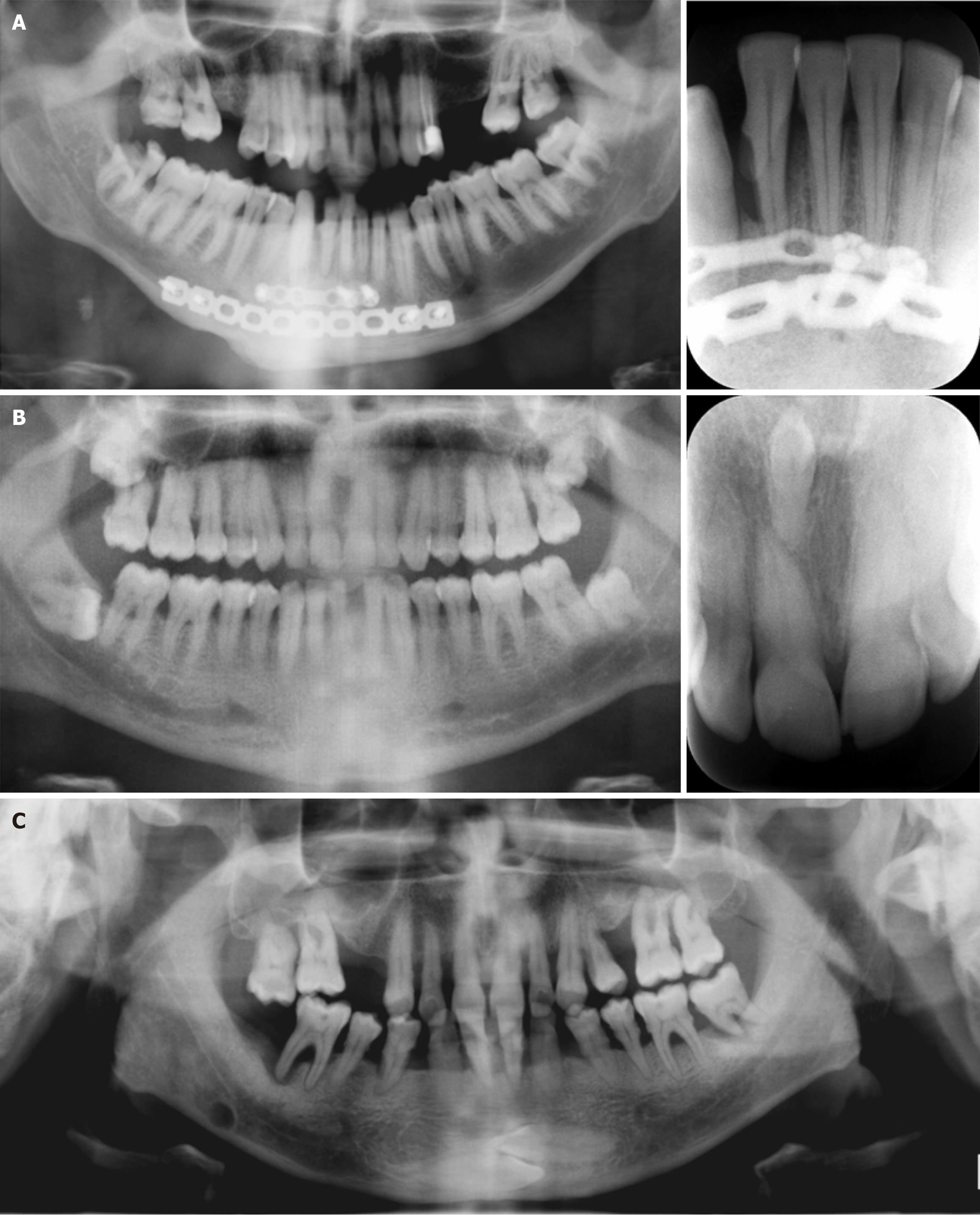
Figure 2 Panoramic images.
A: Patient with fixation screws and no visible alveolar bone defect in the mandibular anterior region on cropped panoramic radiography. Significant bone loss was evident around the right mandibular lateral incisor on cropped periapical radiography; B: A 35-year-old male patient with a vague radiopacity in the maxillary anterior region on cropped panoramic radiography. A clearly depicted inverted mesiodens tooth was evident between the maxillary incisors on cropped periapical radiography; C: Cropped panoramic image of a 55-year-old male patient with aggressive periodontitis and two horizontally impacted canine teeth visible on the mandibular anterior region. Also, a well-defined round Stafne bone cavity can be seen on the right mandibular region extending under the mandibular canal.
- Citation: Kamburoğlu K. Trends in dentomaxillofacial radiology. World J Radiol 2025; 17(1): 97255
- URL: https://www.wjgnet.com/1949-8470/full/v17/i1/97255.htm
- DOI: https://dx.doi.org/10.4329/wjr.v17.i1.97255


