Copyright
©The Author(s) 2025.
World J Radiol. Jan 28, 2025; 17(1): 100794
Published online Jan 28, 2025. doi: 10.4329/wjr.v17.i1.100794
Published online Jan 28, 2025. doi: 10.4329/wjr.v17.i1.100794
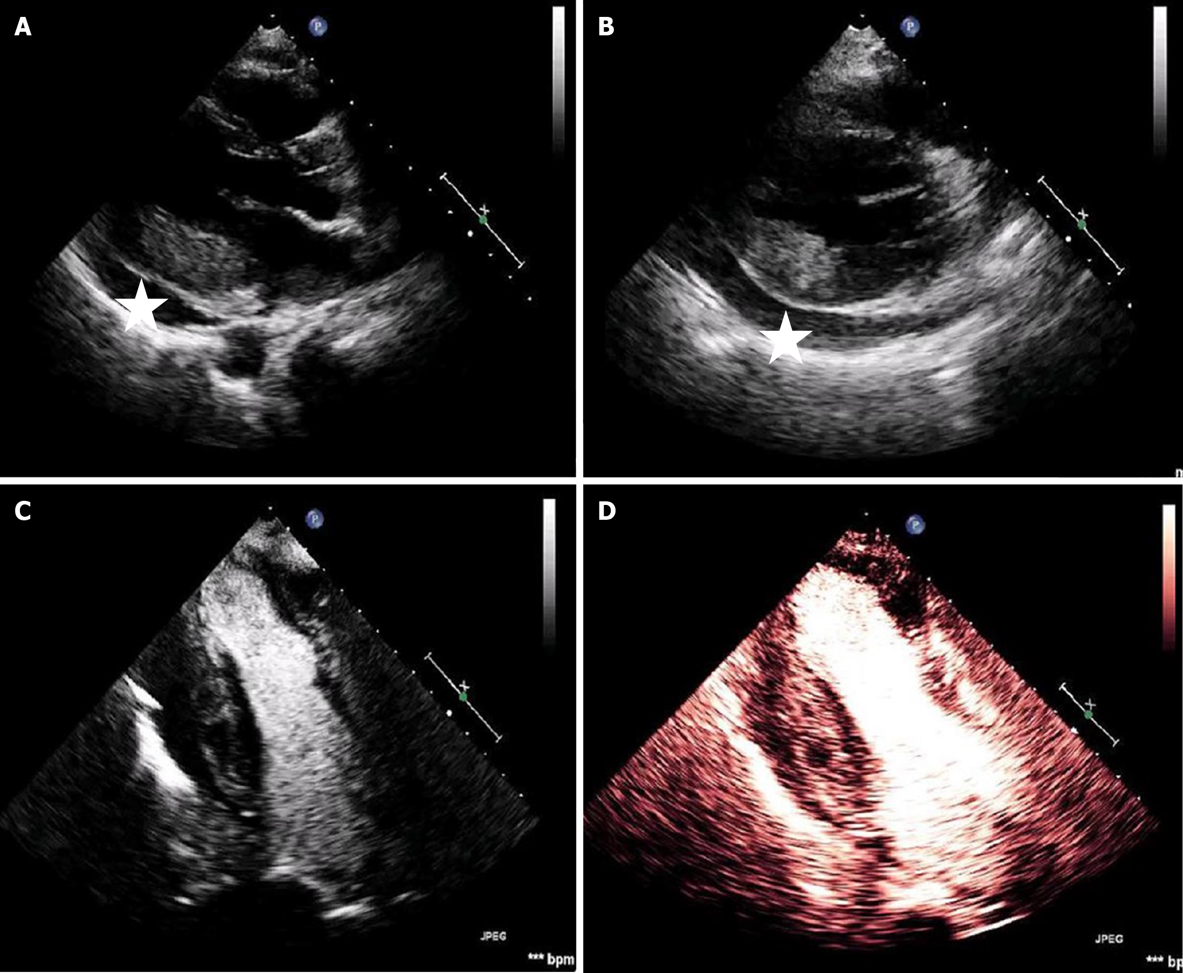
Figure 3 Echocardiography study.
A: In the parasternal long axis view increased thickness of the inferior wall when compared to the anterospetal wall can be noted. There is also pericardial effusion (asterisk); B: Short axis view; C and D: Apical views using contrast agents. There is perfusion inhomogeneity of the inferior wall.
- Citation: Latsios G, Dimitroglou Y, Lazaros G, Alexopoulos N, Tolis I, Aggeli C, Tsioufis C. Differentiating between immune checkpoint inhibitor-induced myocarditis and cardiac metastasis in a cardio-oncology patient presenting with myocardial infarction: A case report. World J Radiol 2025; 17(1): 100794
- URL: https://www.wjgnet.com/1949-8470/full/v17/i1/100794.htm
- DOI: https://dx.doi.org/10.4329/wjr.v17.i1.100794


