Copyright
©The Author(s) 2024.
World J Radiol. Sep 28, 2024; 16(9): 489-496
Published online Sep 28, 2024. doi: 10.4329/wjr.v16.i9.489
Published online Sep 28, 2024. doi: 10.4329/wjr.v16.i9.489
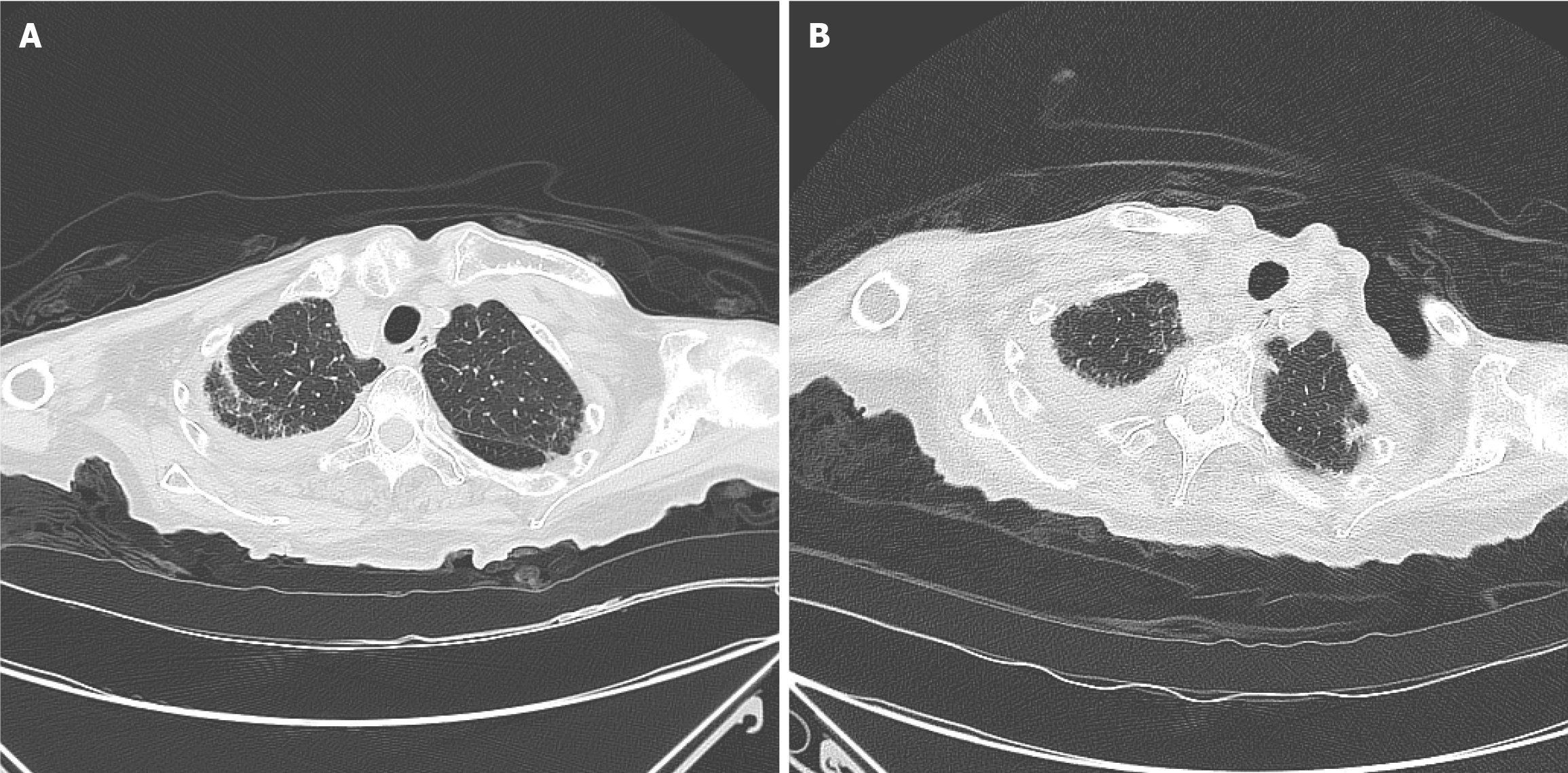
Figure 2 Comparison of lung computed tomography findings.
A: Chest computed tomography (CT) findings upon admission on December 29, 2022: Infectious lesions were present in both lungs with a small amount of fluid on both sides, mainly on the right side; B: Chest CT findings before the patient was discharged on January 6, 2023.
- Citation: Zhang YX, Tang J, Zhu D, Wu CY, Liang ML, Huang YT. Prolonged course of Paxlovid administration in a centenarian with COVID-19: A case report. World J Radiol 2024; 16(9): 489-496
- URL: https://www.wjgnet.com/1949-8470/full/v16/i9/489.htm
- DOI: https://dx.doi.org/10.4329/wjr.v16.i9.489


