Copyright
©The Author(s) 2024.
World J Radiol. Sep 28, 2024; 16(9): 473-481
Published online Sep 28, 2024. doi: 10.4329/wjr.v16.i9.473
Published online Sep 28, 2024. doi: 10.4329/wjr.v16.i9.473
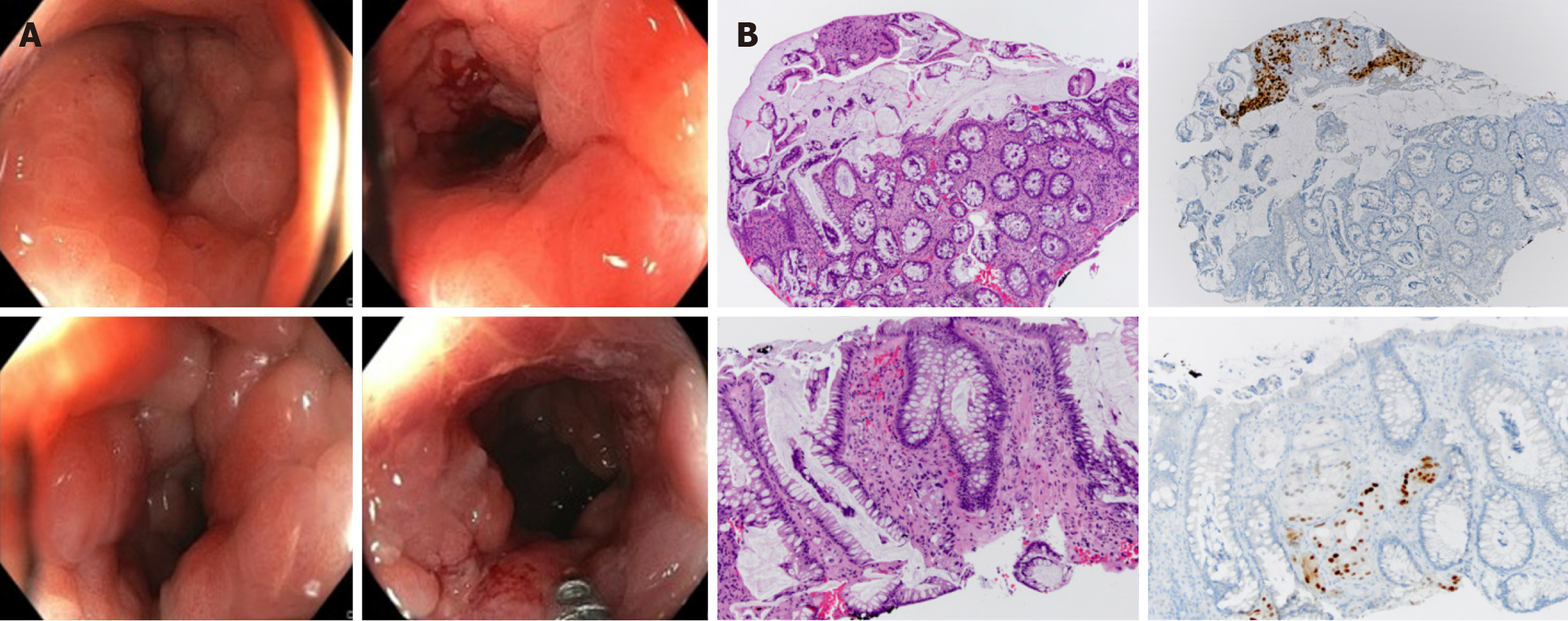
Figure 9 Colonoscopy and histological images of case 1 demostrating advanced metastatic prostate adenocarcinoma.
A: Colonoscopy image showing a reduced rectal lumen with congestive mucosa exhibiting increased consistency and loss of elasticity; B: Histological images of metastatic prostate adenocarcinoma utilizing Hematoxylin and Eosin staining in conjunction with immunohistochemical evaluation for the NKX marker. Groups of neoplastic cells arranged in nests and tubular structures. These cells exhibited atypical nuclei with prominent nucleoli and were positive for NKX, a highly specific marker for, albeit not exclusively indicative of, prostate origin.
- Citation: Labra AA, Schiappacasse G, Cocio RA, Torres JT, González FO, Cristi JA, Schultz M. Secondary rectal linitis plastica caused by prostatic adenocarcinoma - magnetic resonance imaging findings and dissemination pathways: A case report. World J Radiol 2024; 16(9): 473-481
- URL: https://www.wjgnet.com/1949-8470/full/v16/i9/473.htm
- DOI: https://dx.doi.org/10.4329/wjr.v16.i9.473


