Copyright
©The Author(s) 2024.
World J Radiol. Sep 28, 2024; 16(9): 473-481
Published online Sep 28, 2024. doi: 10.4329/wjr.v16.i9.473
Published online Sep 28, 2024. doi: 10.4329/wjr.v16.i9.473
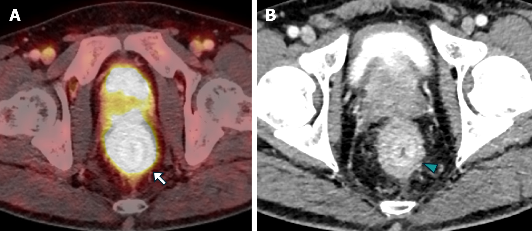
Figure 7 The positron emission tomography/computed tomography PSMA images in case 2.
A and B: Axial FUSION (A) and computed tomography (B) of abdomen and pelvis with intravenous contrast revealed a stratified parietal thickening of the rectal wall, displaying significant uptake of the radiotracer, thus confirming the prostatic origin of the neoplastic infiltration (white arrow). Furthermore, concentric impregnation with intravenous contrast (green arrowhead) was also observed.
- Citation: Labra AA, Schiappacasse G, Cocio RA, Torres JT, González FO, Cristi JA, Schultz M. Secondary rectal linitis plastica caused by prostatic adenocarcinoma - magnetic resonance imaging findings and dissemination pathways: A case report. World J Radiol 2024; 16(9): 473-481
- URL: https://www.wjgnet.com/1949-8470/full/v16/i9/473.htm
- DOI: https://dx.doi.org/10.4329/wjr.v16.i9.473


