Copyright
©The Author(s) 2024.
World J Radiol. Sep 28, 2024; 16(9): 473-481
Published online Sep 28, 2024. doi: 10.4329/wjr.v16.i9.473
Published online Sep 28, 2024. doi: 10.4329/wjr.v16.i9.473
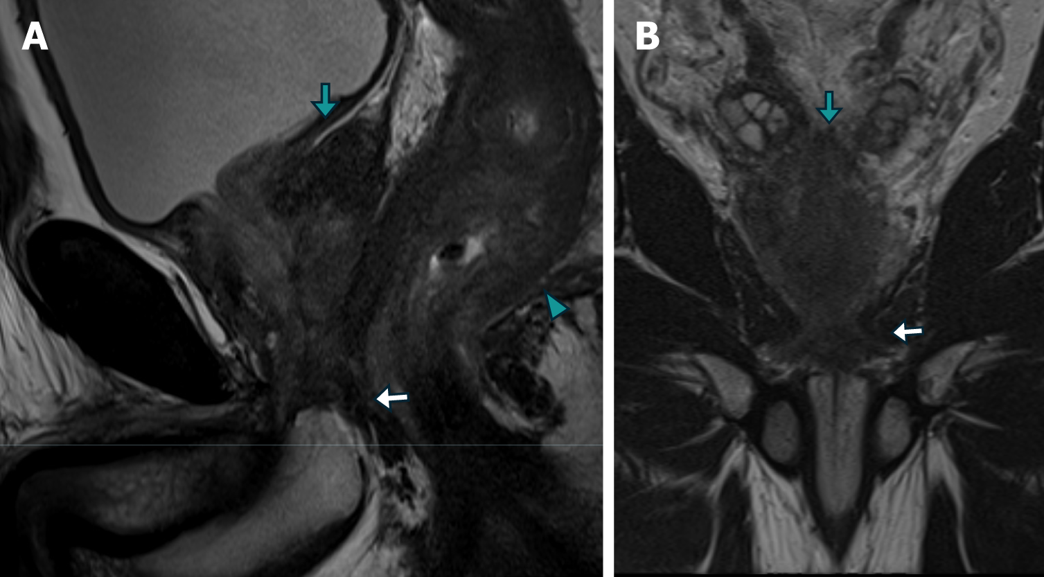
Figure 5 Magnetic resonance imaging images of case 2 demonstrating metastatic prostate adenocarcinoma.
A and B: The sagittal (A) and Coronal (B) T2-weighted turbo spin echo images demonstrate diffuse neoplastic involvement of the prostatic parenchyma, with extension to the rectal wall (arrowhead), base of the seminal vesicles (green arrow), and external urethral sphincter (white arrow).
- Citation: Labra AA, Schiappacasse G, Cocio RA, Torres JT, González FO, Cristi JA, Schultz M. Secondary rectal linitis plastica caused by prostatic adenocarcinoma - magnetic resonance imaging findings and dissemination pathways: A case report. World J Radiol 2024; 16(9): 473-481
- URL: https://www.wjgnet.com/1949-8470/full/v16/i9/473.htm
- DOI: https://dx.doi.org/10.4329/wjr.v16.i9.473


