Copyright
©The Author(s) 2024.
World J Radiol. Sep 28, 2024; 16(9): 473-481
Published online Sep 28, 2024. doi: 10.4329/wjr.v16.i9.473
Published online Sep 28, 2024. doi: 10.4329/wjr.v16.i9.473
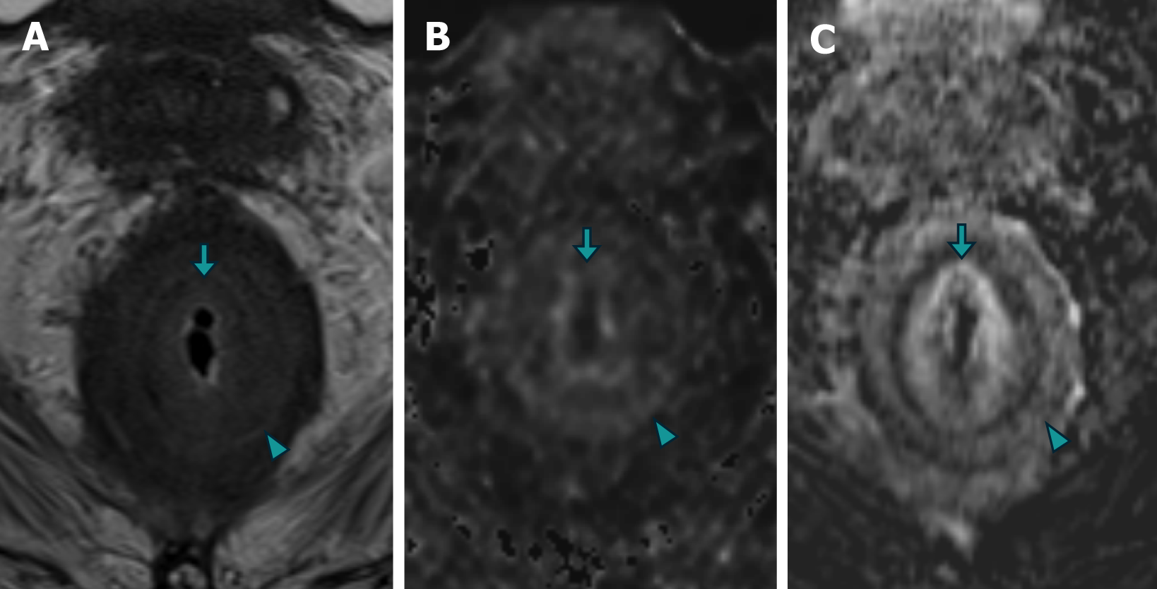
Figure 4 Magnetic resonance imaging images of case 1 demonstrating concentric "target-like" involvement of the rectal parietal wall due to secondary linitis plastica.
A: The axial images obtained from T2 turbo spin echo sequence; B: Diffusion-weighted imaging (DWI); C: T1 gradient recalled echo volumetric interpolated breath-hold examination with contrast-enhanced subtraction technique revealed a stratified parietal thickening of the rectal wall. This thickening displayed concentric areas of intermediate signal intensity on T2-weighted images, restricted diffusion on DWI, and enhancement on contrast-enhanced images, affecting both the submucosal (arrow) and muscular (arrowhead) layers.
- Citation: Labra AA, Schiappacasse G, Cocio RA, Torres JT, González FO, Cristi JA, Schultz M. Secondary rectal linitis plastica caused by prostatic adenocarcinoma - magnetic resonance imaging findings and dissemination pathways: A case report. World J Radiol 2024; 16(9): 473-481
- URL: https://www.wjgnet.com/1949-8470/full/v16/i9/473.htm
- DOI: https://dx.doi.org/10.4329/wjr.v16.i9.473


