Copyright
©The Author(s) 2024.
World J Radiol. Sep 28, 2024; 16(9): 466-472
Published online Sep 28, 2024. doi: 10.4329/wjr.v16.i9.466
Published online Sep 28, 2024. doi: 10.4329/wjr.v16.i9.466
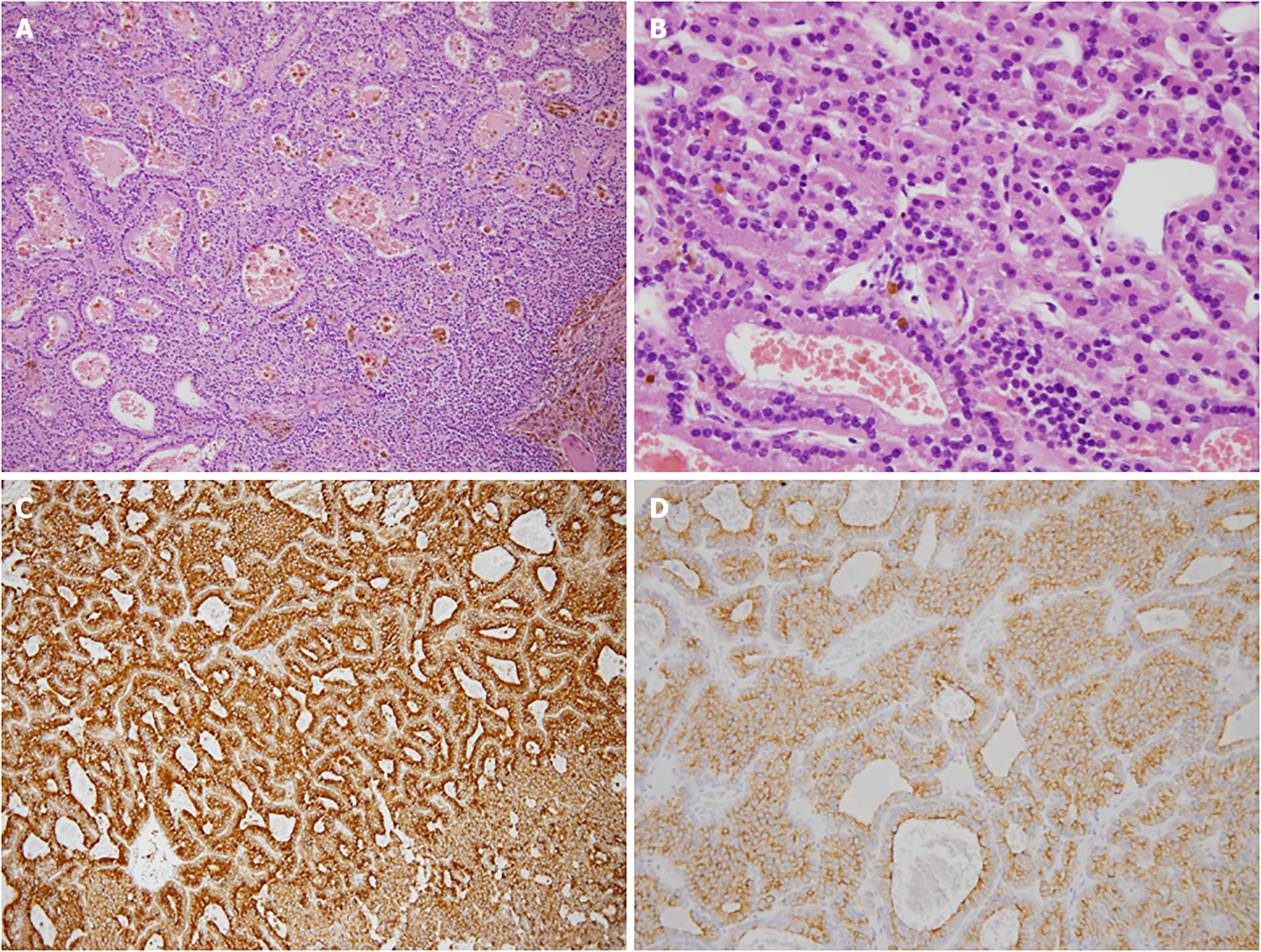
Figure 3 Histology of the nodules.
A: Hematoxylin and eosin (HE) stain, magnification 100 ×. Endocrine cells with blood are visible. Monotonous cells with round nuclei, occasional small nucleoli and eosinophilic cytoplasm arranged in follicular and landular patterns are apparent; B: HE stain, magnification 400 ×. Homogeneous nucleus and no obvious mitoses are shown; C: Parathyroid hormone immunostain, magnification 400 ×; D: Chromogranin A stain, magnification 400 ×.
- Citation: Chiang PH, Ko KH, Peng YJ, Huang TW, Tang SE. Hyperparathyroidism presented as multiple pulmonary nodules in hemodialysis patient status post parathyroidectomy: A case report. World J Radiol 2024; 16(9): 466-472
- URL: https://www.wjgnet.com/1949-8470/full/v16/i9/466.htm
- DOI: https://dx.doi.org/10.4329/wjr.v16.i9.466


