Copyright
©The Author(s) 2024.
World J Radiol. Sep 28, 2024; 16(9): 466-472
Published online Sep 28, 2024. doi: 10.4329/wjr.v16.i9.466
Published online Sep 28, 2024. doi: 10.4329/wjr.v16.i9.466
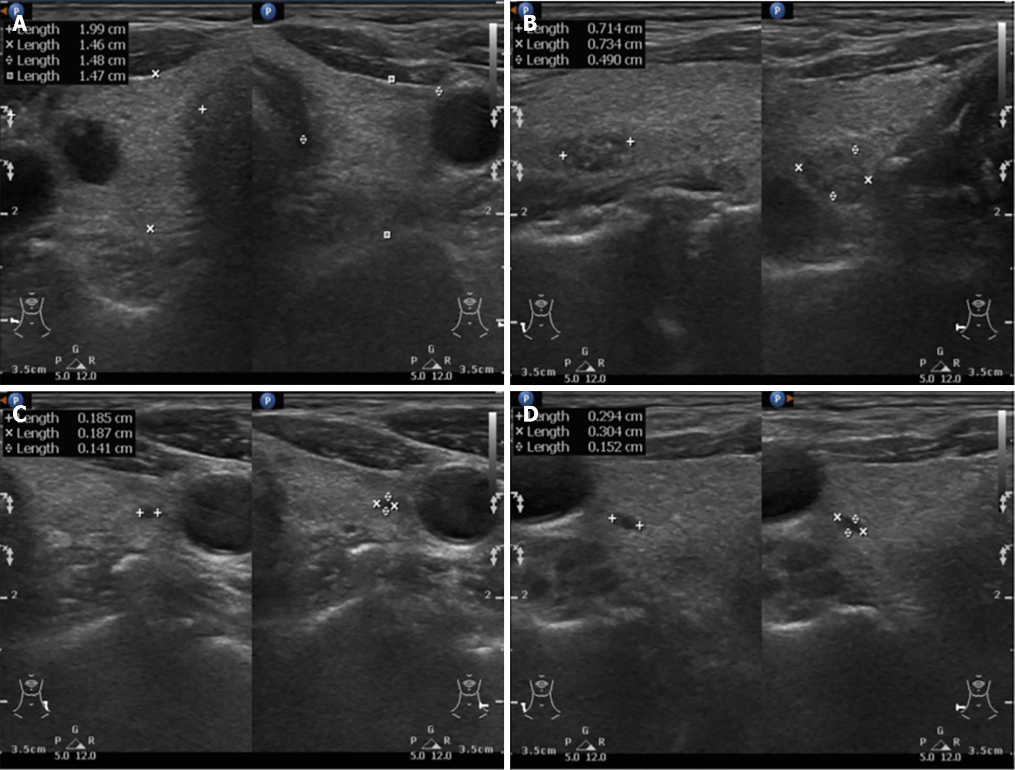
Figure 2 Neck sonography shows right and left lobe thyroid and multinodular goiter.
A: Right and left lobe thyroid; B: Right side nodular goiter 0.73 cm × 0.49 cm × 0.71 cm; C: Left side nodular goiter 0.18 cm × 0.14 cm × 0.18 cm; D: Right side nodular goiter 0.30 cm × 0.15 cm × 0.29 cm.
- Citation: Chiang PH, Ko KH, Peng YJ, Huang TW, Tang SE. Hyperparathyroidism presented as multiple pulmonary nodules in hemodialysis patient status post parathyroidectomy: A case report. World J Radiol 2024; 16(9): 466-472
- URL: https://www.wjgnet.com/1949-8470/full/v16/i9/466.htm
- DOI: https://dx.doi.org/10.4329/wjr.v16.i9.466


