Copyright
©The Author(s) 2024.
World J Radiol. Sep 28, 2024; 16(9): 453-459
Published online Sep 28, 2024. doi: 10.4329/wjr.v16.i9.453
Published online Sep 28, 2024. doi: 10.4329/wjr.v16.i9.453
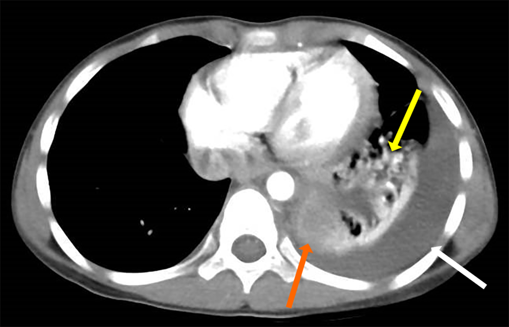
Figure 1 Exemplar chest contrast-enhanced computed tomography results in case 2 (a 7-year-old boy), showing an approximately 52.
9 mm × 40.5 mm × 40.8 mm-sized oval lesion at the level of the 9th-10th thoracic vertebra (orange arrow), with a pleural effusion (white arrow) and pneumonia (yellow arrow). No obvious enhancement was observed.
- Citation: Jiang MY, Wang YX, Lu ZW, Zheng YJ. Extralobar pulmonary sequestration in children with abdominal pain: Four case reports. World J Radiol 2024; 16(9): 453-459
- URL: https://www.wjgnet.com/1949-8470/full/v16/i9/453.htm
- DOI: https://dx.doi.org/10.4329/wjr.v16.i9.453


