Copyright
©The Author(s) 2024.
World J Radiol. Sep 28, 2024; 16(9): 439-445
Published online Sep 28, 2024. doi: 10.4329/wjr.v16.i9.439
Published online Sep 28, 2024. doi: 10.4329/wjr.v16.i9.439
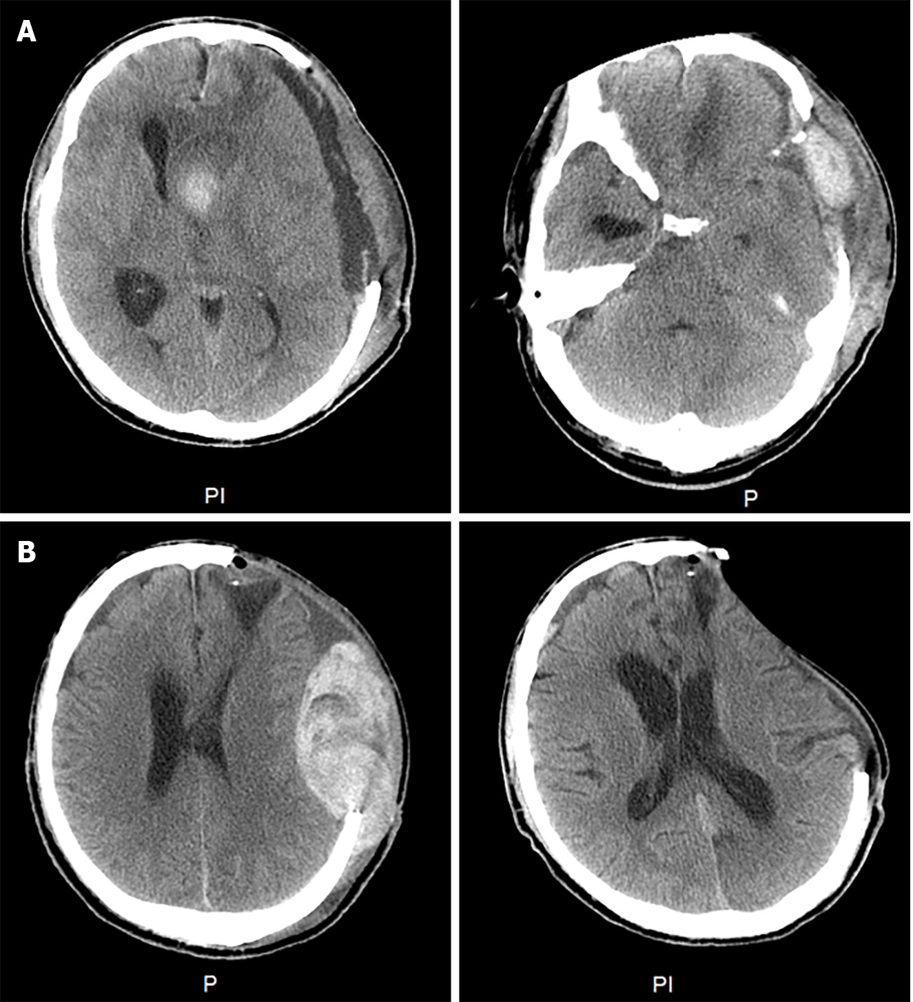
Figure 1 Head computed tomography scan.
A: Head computed tomography scan at the time of initial treatment. The left side is a preoperative image. The right side is head computed tomography scan at rebleeding on day 4; B: Head computed tomography scan during treatment. The left side is a preoperative image. The right side is a postoperative image.
- Citation: Wang L, Zhang N, Liang DC, Zhang HL, Lin LQ. Acquired factor XIII deficiency presenting with multiple intracranial hemorrhages and right hip hematoma: A case report. World J Radiol 2024; 16(9): 439-445
- URL: https://www.wjgnet.com/1949-8470/full/v16/i9/439.htm
- DOI: https://dx.doi.org/10.4329/wjr.v16.i9.439


