Copyright
©The Author(s) 2024.
World J Radiol. Sep 28, 2024; 16(9): 398-406
Published online Sep 28, 2024. doi: 10.4329/wjr.v16.i9.398
Published online Sep 28, 2024. doi: 10.4329/wjr.v16.i9.398
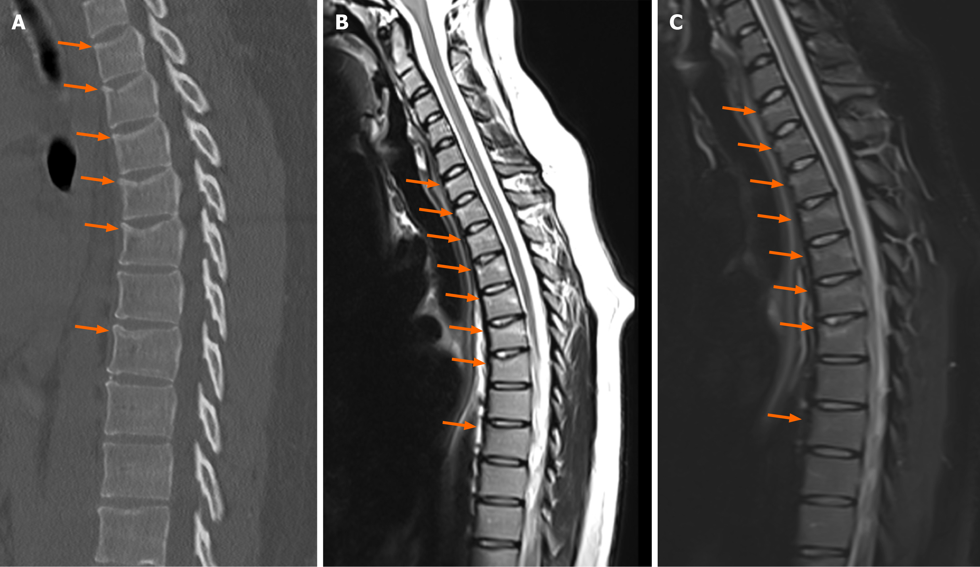
Figure 2 Compression fracture.
A 27-year old male who was injured while escaping from the earthquake. A: Computed tomography image reveals T1-T5 vertebrae and T7 vertebra compression fractures (arrows); B and C: T2-STIR and T2W images show C6-T5 vertebrae and T7 vertebra compression fractures (arrows) and edema around them.
- Citation: Bolukçu A, Erdemir AG, İdilman İS, Yildiz AE, Çoban Çifçi G, Onur MR, Akpinar E. Radiological findings of February 2023 twin earthquakes-related spine injuries. World J Radiol 2024; 16(9): 398-406
- URL: https://www.wjgnet.com/1949-8470/full/v16/i9/398.htm
- DOI: https://dx.doi.org/10.4329/wjr.v16.i9.398


