Copyright
©The Author(s) 2024.
World J Radiol. Sep 28, 2024; 16(9): 398-406
Published online Sep 28, 2024. doi: 10.4329/wjr.v16.i9.398
Published online Sep 28, 2024. doi: 10.4329/wjr.v16.i9.398
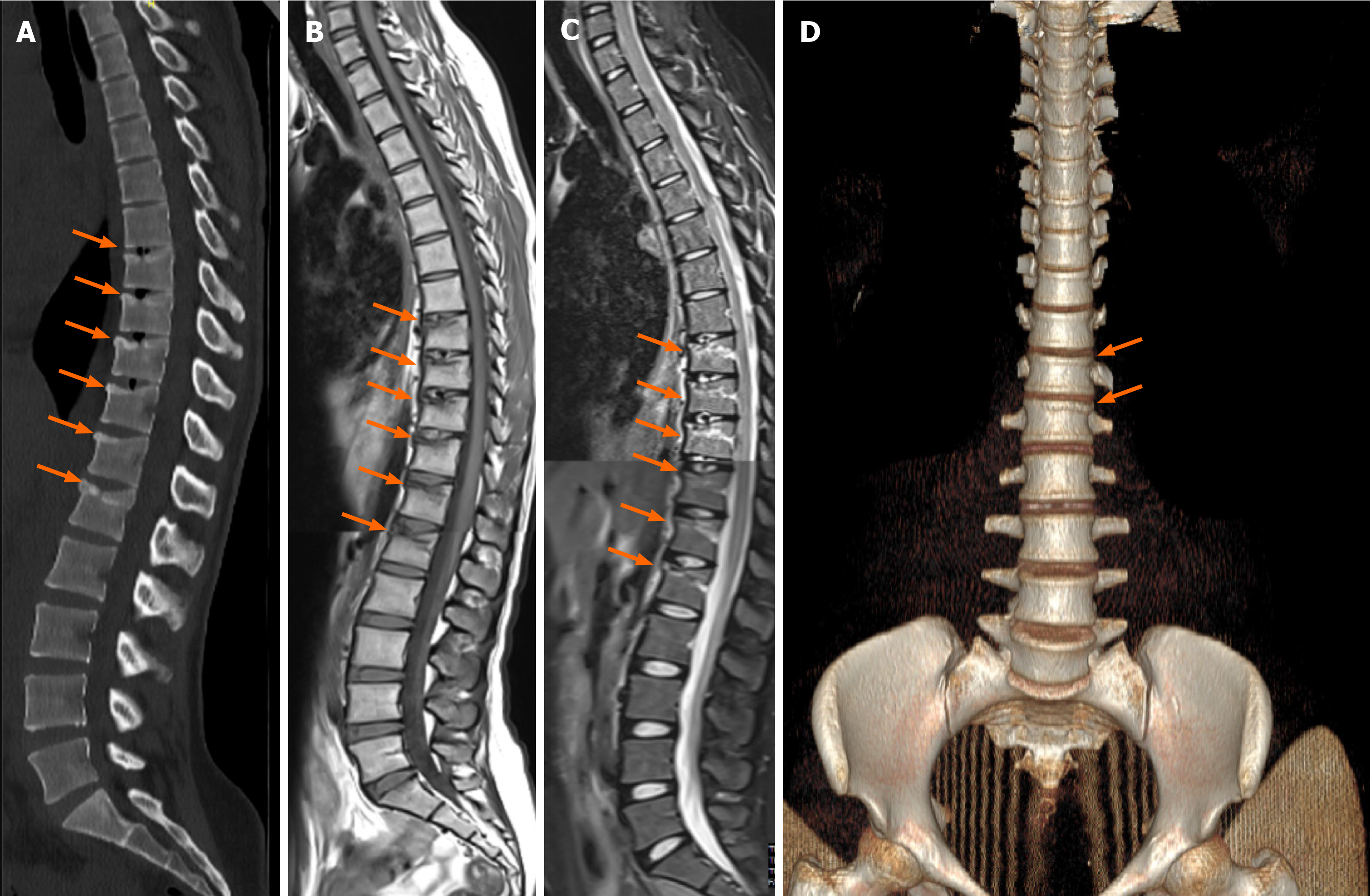
Figure 1 Prolonged fetal posture injury.
A 23-year old male who jammed under debris about 15 minutes. A: Computed tomography image reveals T8 to L1 consecutive mild compression fractures (arrows), which corresponds to thoracolumbar area injury; B and C: T1W image and T2-STIR sequence show T8 to L1 vertebrae fracture and edema around them; D: The image of the volume rendering technique shows especially the fractures of the T12 and L1 vertebrae.
- Citation: Bolukçu A, Erdemir AG, İdilman İS, Yildiz AE, Çoban Çifçi G, Onur MR, Akpinar E. Radiological findings of February 2023 twin earthquakes-related spine injuries. World J Radiol 2024; 16(9): 398-406
- URL: https://www.wjgnet.com/1949-8470/full/v16/i9/398.htm
- DOI: https://dx.doi.org/10.4329/wjr.v16.i9.398


