Copyright
©The Author(s) 2024.
World J Radiol. Sep 28, 2024; 16(9): 389-397
Published online Sep 28, 2024. doi: 10.4329/wjr.v16.i9.389
Published online Sep 28, 2024. doi: 10.4329/wjr.v16.i9.389
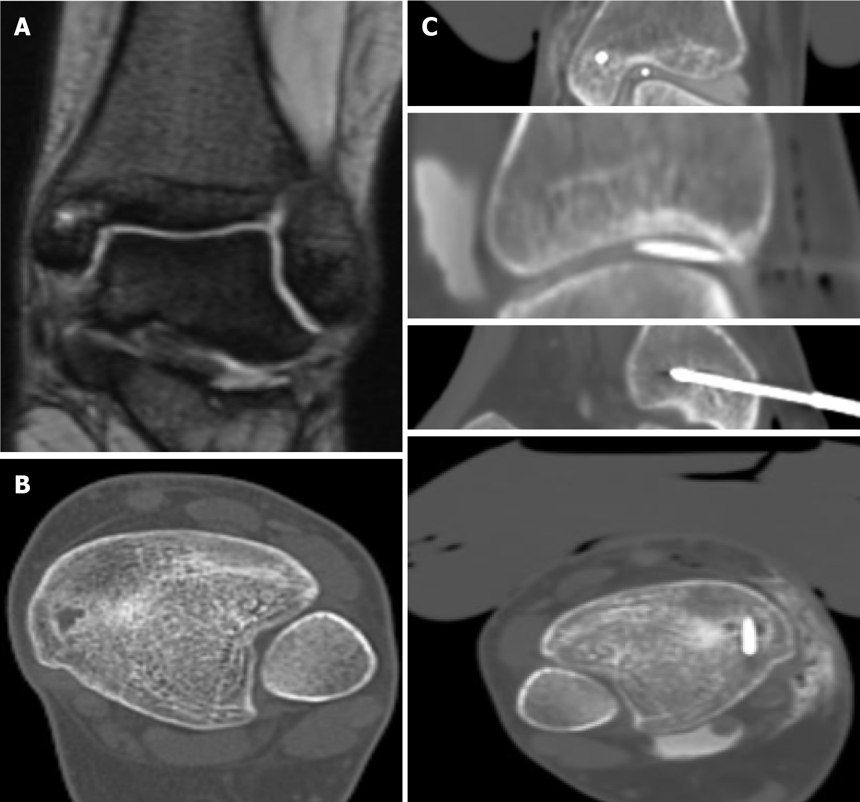
Figure 1 Eighteen-year-old female-osteoid osteoma of the medial malleolus (tibia).
A: Coronal T2WI image reveals 1 cm lesion of the medial malleous in close proximity to the skin and the ankle joint; B: Computed tomography image demonstrates a lytic lesion with reactive sclerosis. The lesion was biopsied and confirmed as an osteoid osteoma; C: Cryoablation procedure: Cryoprobe placement inside the nidus and a spinal needle in the joint for active warming during the procedure. Skin was hydrodissected with a mixture of saline and local anesthetic and a warm glove was applied for protection.
- Citation: Michailidis A, Panos A, Samoladas E, Dimou G, Mingou G, Kosmoliaptsis P, Arvaniti M, Giankoulof C, Petsatodis E. Cryoablation of osteoid osteomas: Is it a valid treatment option? World J Radiol 2024; 16(9): 389-397
- URL: https://www.wjgnet.com/1949-8470/full/v16/i9/389.htm
- DOI: https://dx.doi.org/10.4329/wjr.v16.i9.389


