Copyright
©The Author(s) 2024.
World J Radiol. Sep 28, 2024; 16(9): 380-388
Published online Sep 28, 2024. doi: 10.4329/wjr.v16.i9.380
Published online Sep 28, 2024. doi: 10.4329/wjr.v16.i9.380
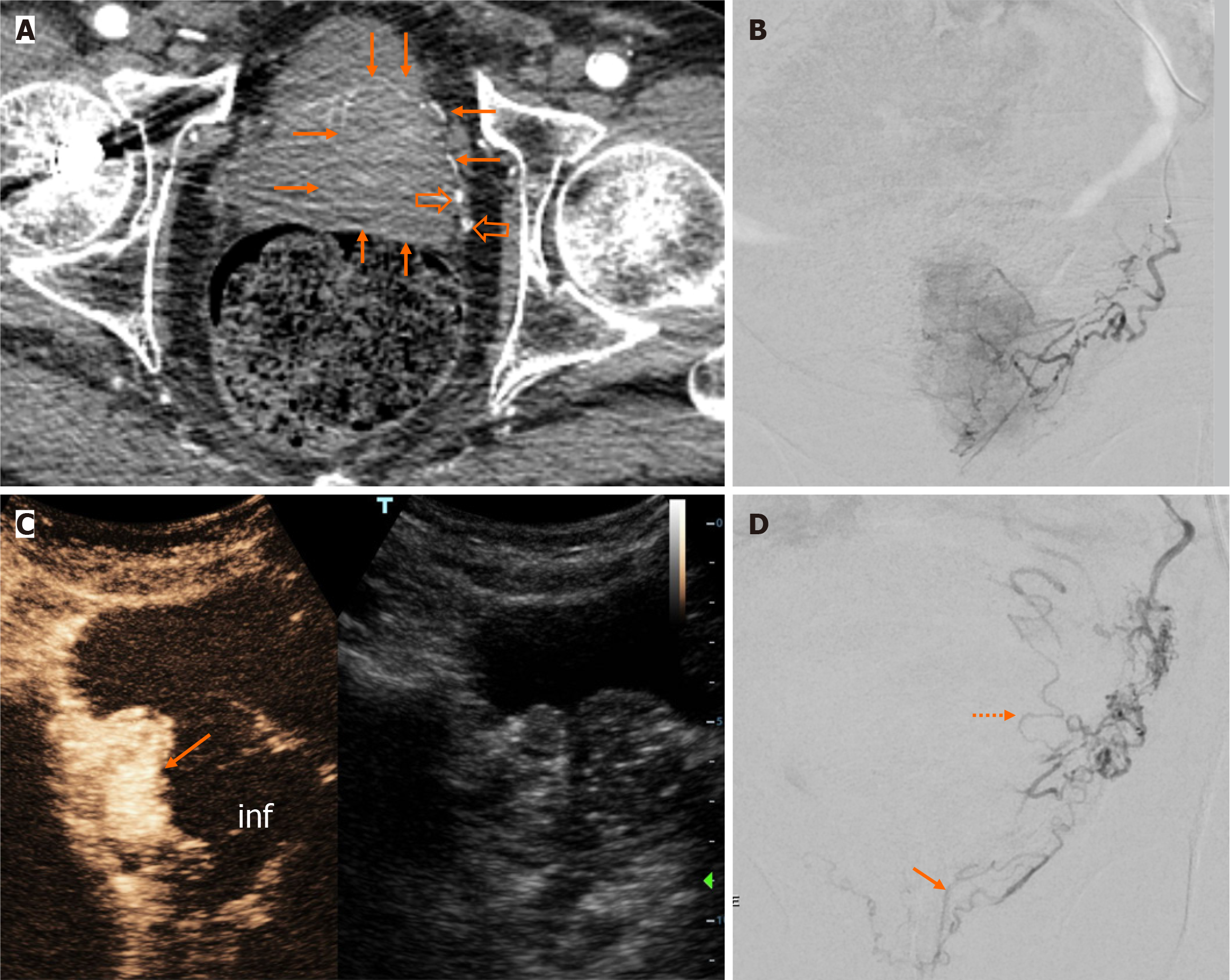
Figure 2 Representative images from a clinically successful case of intentionally unilateral prostatic artery embolization (subgroup B).
A: Axial computed tomographic angiography image shows a dominant left prostatic lobe (arrows). Left prostatic artery (PA) branches (empty arrows) are also prominent compared to the right side; B: Left PA angiogram shows dense blush of the ipsilateral prostatic lobe; C: Dual image from intraprocedural contrast-enhanced ultrasonography (contrast-enhanced image on the left, unenhanced, reference B-mode image on the right) 2 minutes post embolization, shows almost complete absence of enhancement of the left lobe (inf) and persistent enhancement of the right lobe (arrow); D: Post embolization angiogram with microcatheter position near the origin of the left PA shows disappearance of the left prostatic blush and activation of anastomoses with rectal (arrow) and vesical (dotted arrow) branches.
- Citation: Moschouris H, Stamatiou K. Intentionally unilateral prostatic artery embolization: Patient selection, technique and potential benefits. World J Radiol 2024; 16(9): 380-388
- URL: https://www.wjgnet.com/1949-8470/full/v16/i9/380.htm
- DOI: https://dx.doi.org/10.4329/wjr.v16.i9.380


