Copyright
©The Author(s) 2024.
World J Radiol. Aug 28, 2024; 16(8): 371-374
Published online Aug 28, 2024. doi: 10.4329/wjr.v16.i8.371
Published online Aug 28, 2024. doi: 10.4329/wjr.v16.i8.371
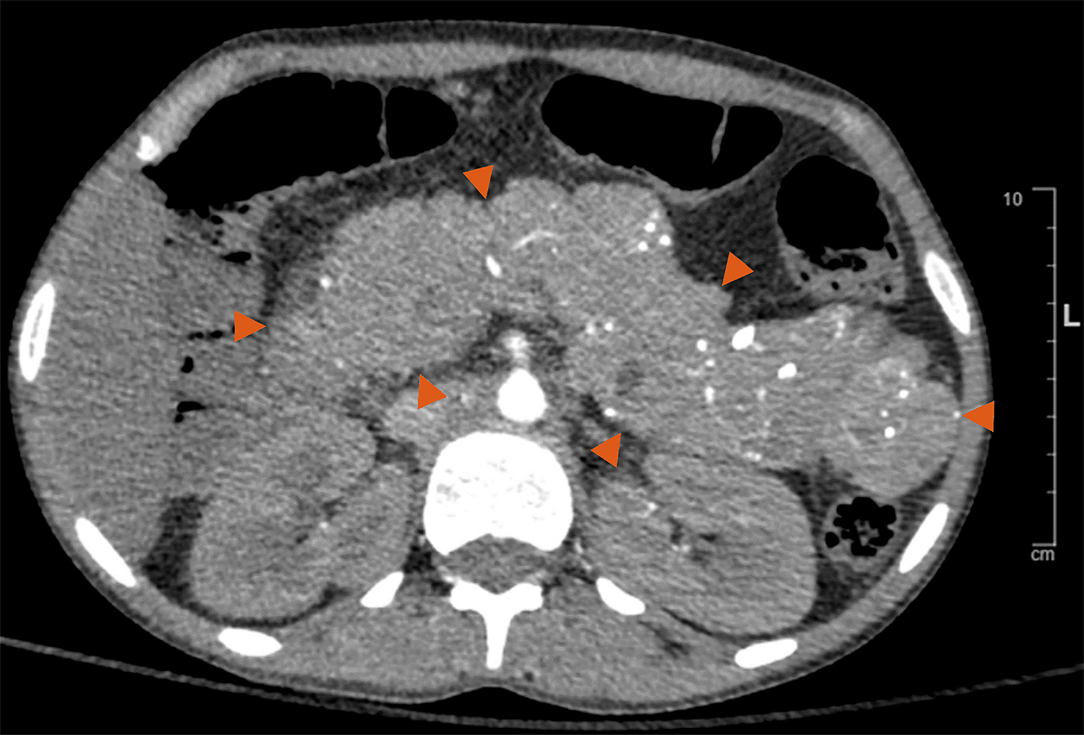
Figure 1 Pancreas in Mahvash disease, axial view of computed tomography with enhancement.
Note the enormously and evenly enlarged pancreas with normal pancreatic contour. Also note the multiple punctate calcifications in the pancreas.
- Citation: Yu R. Plea to radiologists: Please consider Mahvash disease when encountering an enlarged pancreas. World J Radiol 2024; 16(8): 371-374
- URL: https://www.wjgnet.com/1949-8470/full/v16/i8/371.htm
- DOI: https://dx.doi.org/10.4329/wjr.v16.i8.371


