Copyright
©The Author(s) 2024.
World J Radiol. Aug 28, 2024; 16(8): 356-361
Published online Aug 28, 2024. doi: 10.4329/wjr.v16.i8.356
Published online Aug 28, 2024. doi: 10.4329/wjr.v16.i8.356
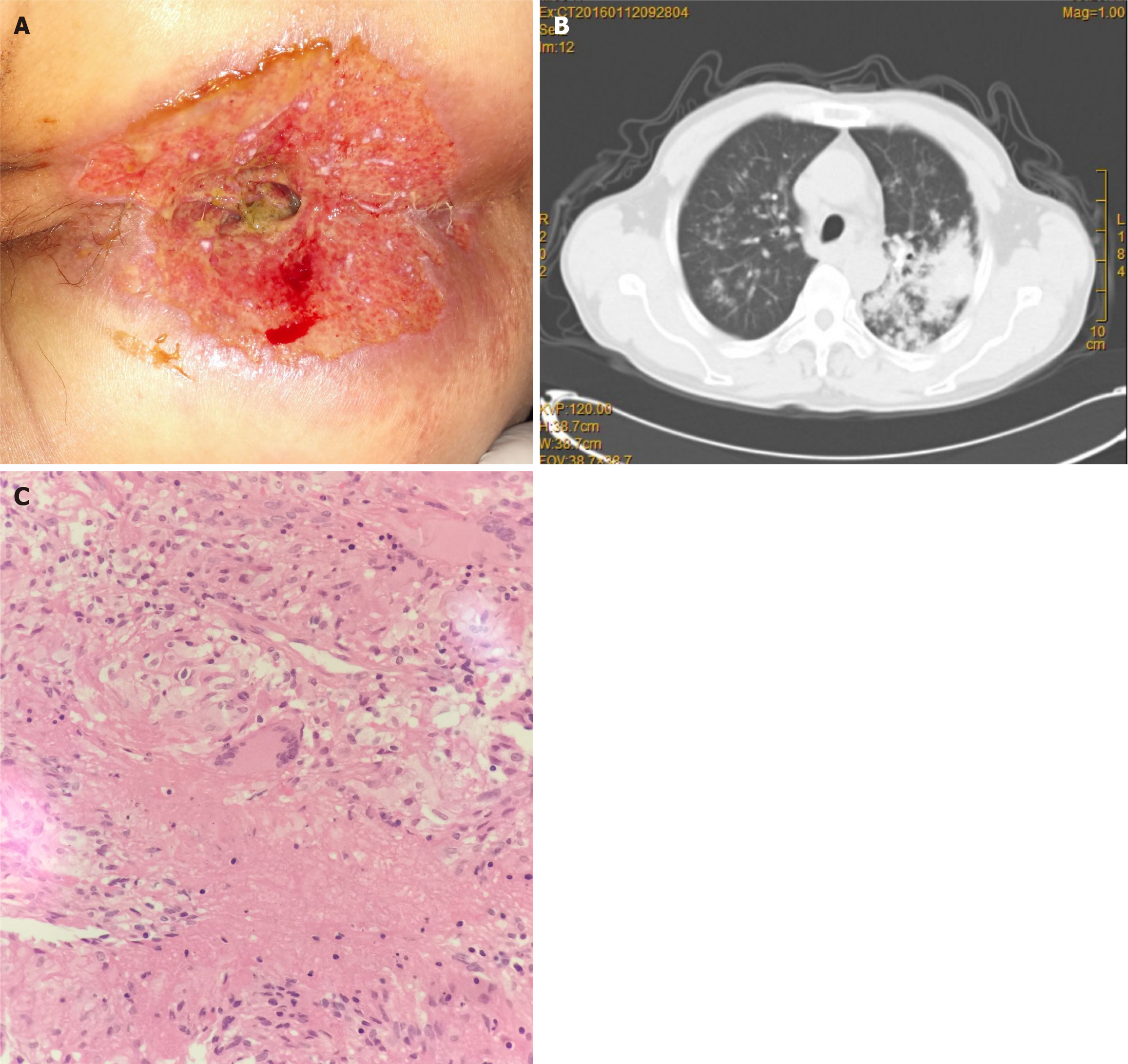
Figure 1 Imaging and macroscopic examination of the perianal tuberculous ulcer.
A: Perianal photographs showing perianal skin ulcers and impaired anal canal integrity (cavity type anus); B: A computed tomography scan of chest pulmonary tuberculosis shows active pulmonary tuberculosis in both lungs; C: A pathological section of the perianal ulcer revealed caseous necrotizing granuloma.
- Citation: Yuan B, Ma CQ. Perianal tuberculous ulcer with active pulmonary, intestinal and orificial tuberculosis: A case report. World J Radiol 2024; 16(8): 356-361
- URL: https://www.wjgnet.com/1949-8470/full/v16/i8/356.htm
- DOI: https://dx.doi.org/10.4329/wjr.v16.i8.356


