Copyright
©The Author(s) 2024.
World J Radiol. Aug 28, 2024; 16(8): 348-355
Published online Aug 28, 2024. doi: 10.4329/wjr.v16.i8.348
Published online Aug 28, 2024. doi: 10.4329/wjr.v16.i8.348
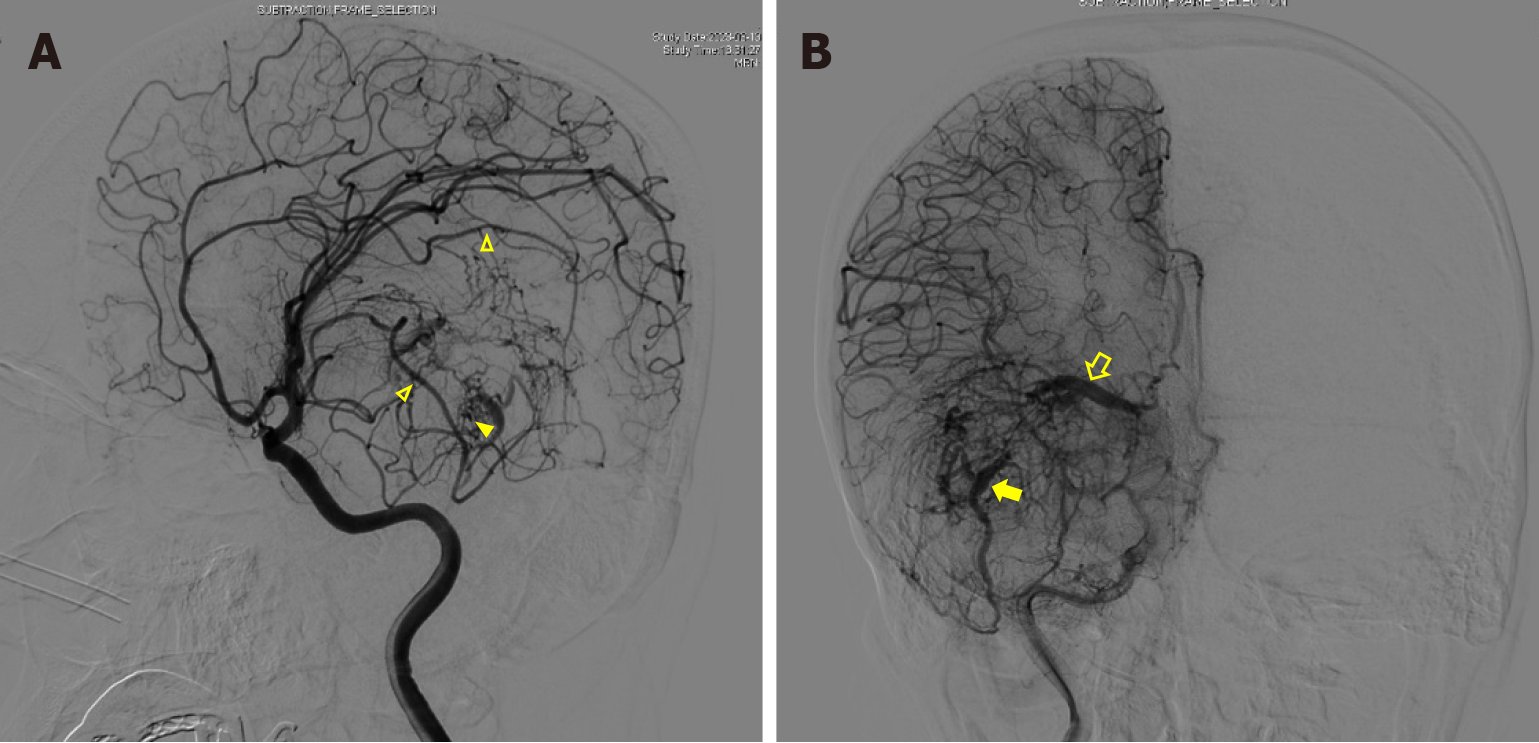
Figure 3 Preoperative cerebral angiography.
A: Two feeding vessels originating from the branches of the right middle artery (open triangles) and two large draining veins were seen in the early arterial phase. Numerous small tortuous blood vessels were observed to form a nidus between the feeding artery and the draining vein (solid triangle); B: In the late arterial phase, both draining veins exhibited a caput medusae distribution consistent with a developmental venous anomaly; one drained into the great cerebral vein and to the sinuses rectus (open arrow), and the other drained through the superior petrosal sinus to the ipsilateral transverse sinus (solid arrow).
- Citation: Guo P, Sun W, Song LX, Cao WY, Li JP. Multimodal imaging for the diagnosis of oligodendroglioma associated with arteriovenous malformation: A case report. World J Radiol 2024; 16(8): 348-355
- URL: https://www.wjgnet.com/1949-8470/full/v16/i8/348.htm
- DOI: https://dx.doi.org/10.4329/wjr.v16.i8.348


