Copyright
©The Author(s) 2024.
World J Radiol. Aug 28, 2024; 16(8): 348-355
Published online Aug 28, 2024. doi: 10.4329/wjr.v16.i8.348
Published online Aug 28, 2024. doi: 10.4329/wjr.v16.i8.348
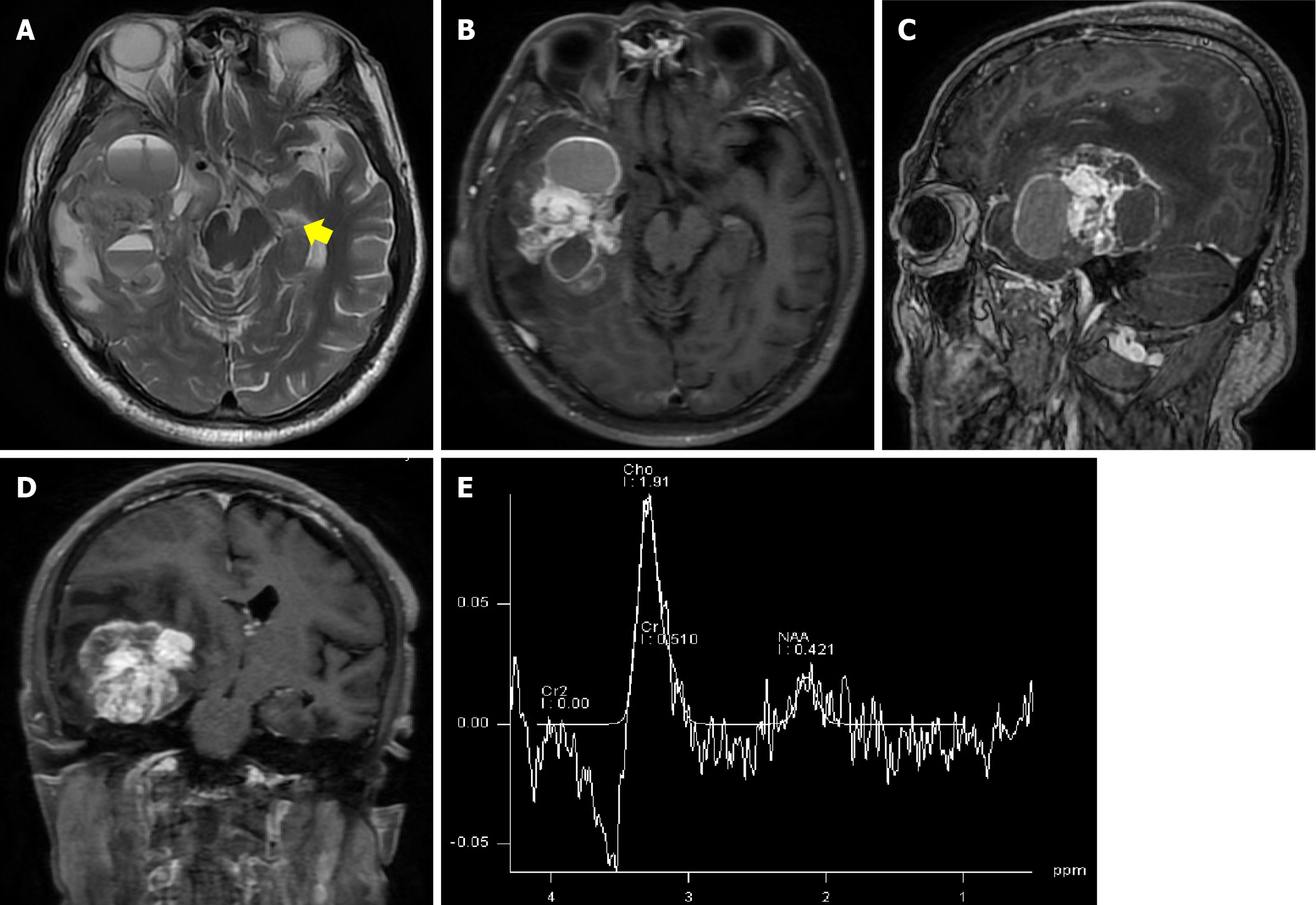
Figure 2 Preoperative neuroimaging.
A: T2-weighted axial magnetic resonance imaging demonstrated cystic and solid space-occupying lesions, a visible air-fluid level in the cyst, and flow void signs within the lesion (arrow); B–D: Gadolinium-enhanced T1-weighted magnetic resonance imaging of the brain revealed a 4.5 cm × 6.3 cm × 4.6 cm necrotic cystic lesion with heterogeneous enhancement involving the basal ganglia, with a midline shift and uncal herniation; E: Magnetic resonance spectroscopy revealed aberrant metabolic function. An increased choline/creatine (Cr) peak ratio and a decreased N-acetyl aspartate/Cr ratio, matching the metabolic signature of glioma, were detected within the lesion.
- Citation: Guo P, Sun W, Song LX, Cao WY, Li JP. Multimodal imaging for the diagnosis of oligodendroglioma associated with arteriovenous malformation: A case report. World J Radiol 2024; 16(8): 348-355
- URL: https://www.wjgnet.com/1949-8470/full/v16/i8/348.htm
- DOI: https://dx.doi.org/10.4329/wjr.v16.i8.348


