Copyright
©The Author(s) 2024.
World J Radiol. Aug 28, 2024; 16(8): 337-347
Published online Aug 28, 2024. doi: 10.4329/wjr.v16.i8.337
Published online Aug 28, 2024. doi: 10.4329/wjr.v16.i8.337
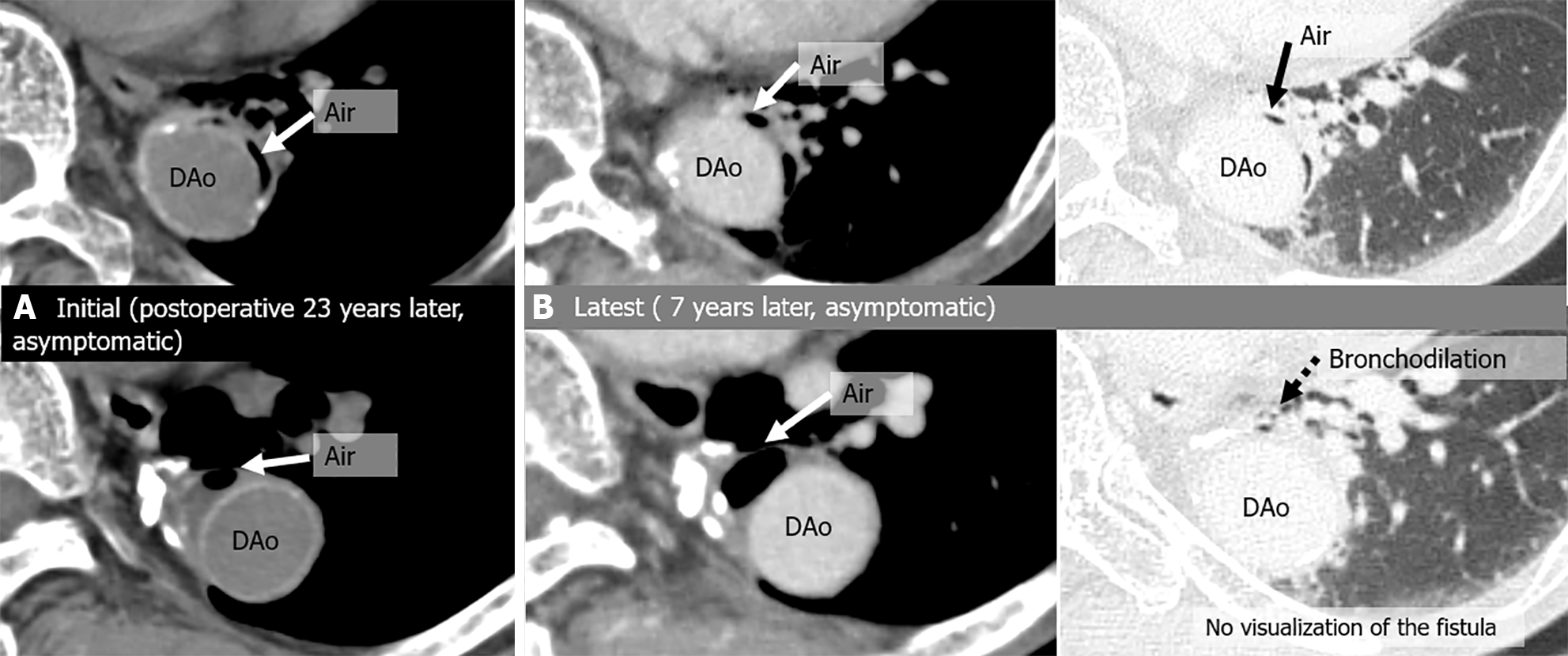
Figure 7 Aortobronchial fistula in an 83-year-old asymptomatic man and a history of descending aorta replacement 23 years earlier for an aneurysm.
A: Initial computed tomography (CT) images show air shadow in the intra-aortic peri-graft space (arrow); B: Latest CT, 7-year follow-up images show residual peri-graft air. A dilated peripheral bronchus is near the graft, but without direct connection with peri-graft air. No severe complications occurred during the 7-year observation period. DAo: Descending aorta.
- Citation: Tsuchiya N, Inafuku H, Yogi S, Iraha Y, Iida G, Ando M, Nagano T, Higa S, Maeda T, Kise Y, Furukawa K, Yonemoto K, Nishie A. Direct visualization of postoperative aortobronchial fistula on computed tomography. World J Radiol 2024; 16(8): 337-347
- URL: https://www.wjgnet.com/1949-8470/full/v16/i8/337.htm
- DOI: https://dx.doi.org/10.4329/wjr.v16.i8.337


