Copyright
©The Author(s) 2024.
World J Radiol. Aug 28, 2024; 16(8): 337-347
Published online Aug 28, 2024. doi: 10.4329/wjr.v16.i8.337
Published online Aug 28, 2024. doi: 10.4329/wjr.v16.i8.337
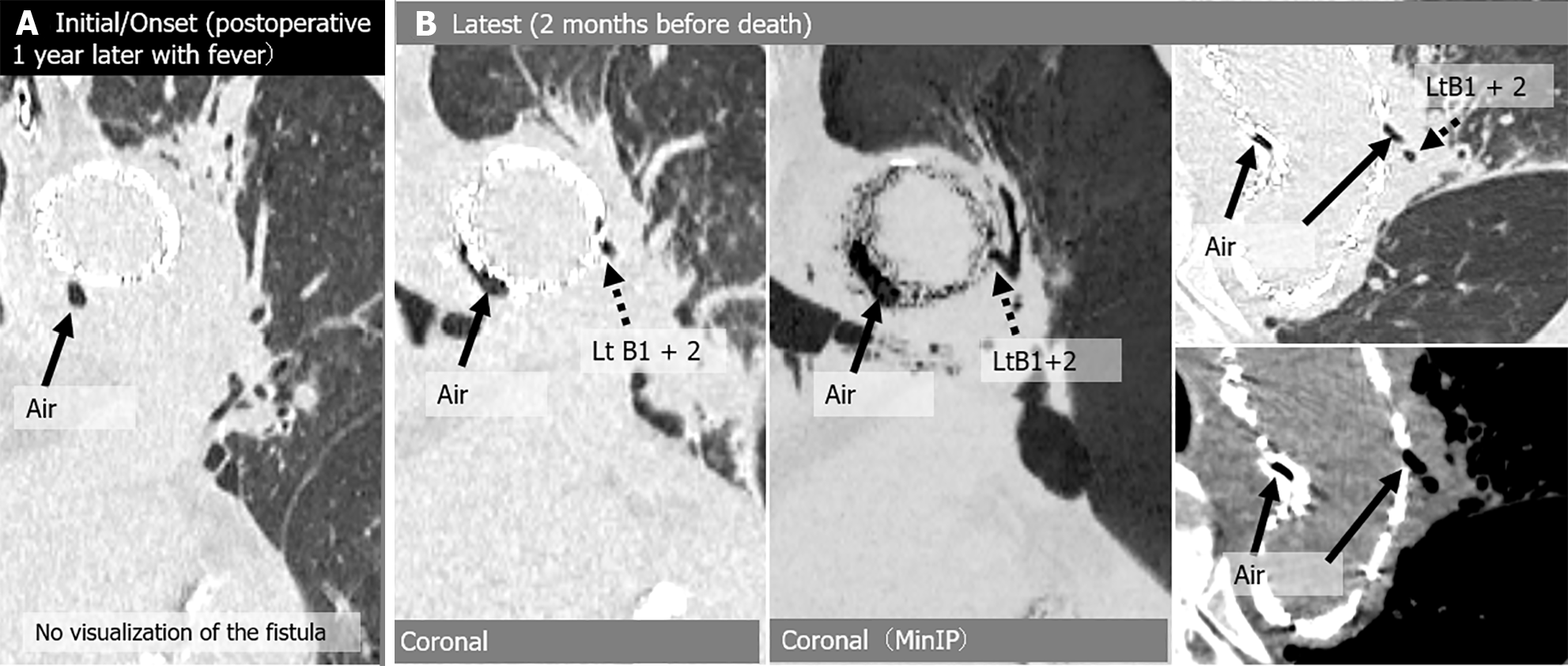
Figure 5 Aortobronchial fistula in a 73-year-old man with fever and a history of thoracic endovascular aortic repair 1 year earlier for infected aneurysm.
One year after the surgery, peri-graft air was first noted when he was diagnosed with a stent graft infection. He was treated with antibiotics, and the infection was brought under control, but a year later the stent graft infection recurred and he died of sepsis. A: Initial/onset computed tomography (CT) images show air shadow in the intra-aortic peri-graft space (arrow), but without direct connection between bronchioles and perigraft air; B: Latest CT images show increased peri-graft air (arrow) and dilated peripheral bronchi (left B1 + 2) communicating with air (arrow). Lt: Left; MinIP: Minimum intensity projection.
- Citation: Tsuchiya N, Inafuku H, Yogi S, Iraha Y, Iida G, Ando M, Nagano T, Higa S, Maeda T, Kise Y, Furukawa K, Yonemoto K, Nishie A. Direct visualization of postoperative aortobronchial fistula on computed tomography. World J Radiol 2024; 16(8): 337-347
- URL: https://www.wjgnet.com/1949-8470/full/v16/i8/337.htm
- DOI: https://dx.doi.org/10.4329/wjr.v16.i8.337


