Copyright
©The Author(s) 2024.
World J Radiol. Aug 28, 2024; 16(8): 337-347
Published online Aug 28, 2024. doi: 10.4329/wjr.v16.i8.337
Published online Aug 28, 2024. doi: 10.4329/wjr.v16.i8.337
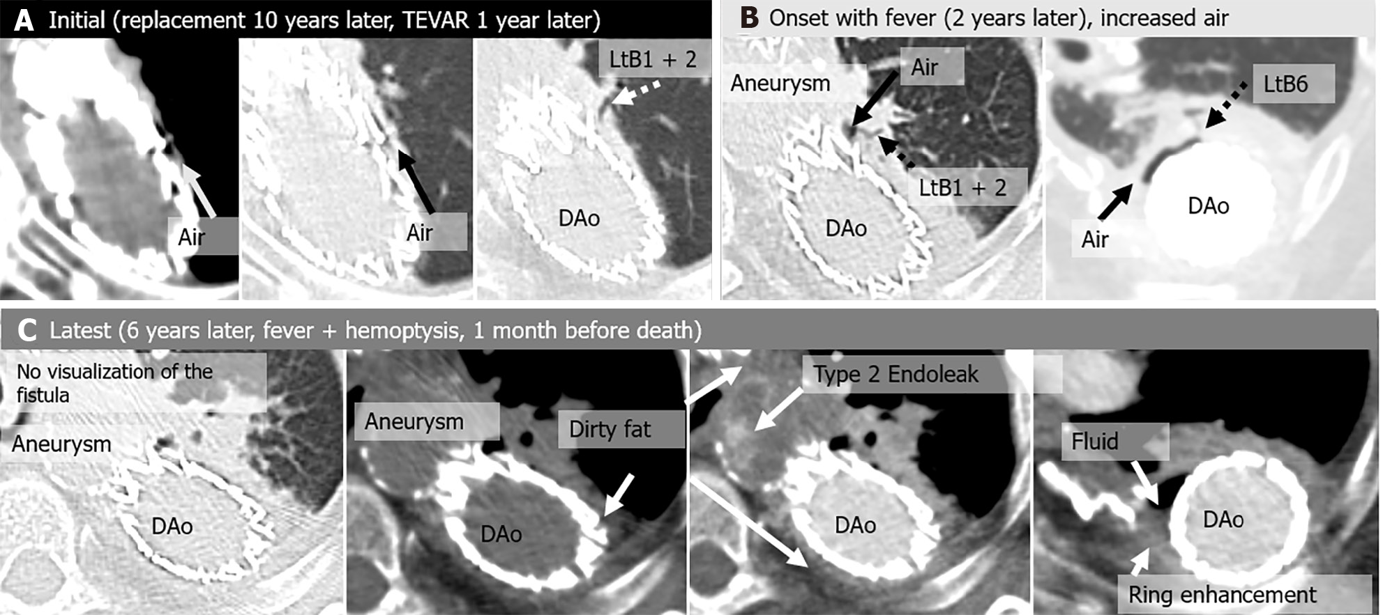
Figure 3 Aortobronchial fistula in an asymptomatic 74-year-old woman, a history of thoracic endovascular aortic repair 1 year earlier for a pseudoaneurysm after replacement of a descending aorta 10 years earlier.
Two years after the initial computed tomography (CT) scan, the patient developed fever and was diagnosed with a stent graft infection. Drainage of the peri-graft abscess was performed, and the infection was controlled, but the infection recurred repeatedly and was complicated by a Type 2 endoleak. At the latest CT scan, the patient had hemoptysis and fever. The patient died from massive hemoptysis 6 years after the initial CT. A: Initial CT images show air shadow in the intra-aortic peri-graft space (arrow) and dilated peripheral bronchi (left B1 + 2) communicating with peri-graft air (arrow); B: Onset CT, 2 years after the initial CT images show dilated peripheral bronchi (left B1 + 2 and B6) communicating with peri-graft air (arrow); C: Latest CT images show peri-graft dirty fat sign and disappearance of direct communication between peripheral bronchi and peri-graft air. CT angiogram demonstrates extravasation of contrast material into the native aneurysm, representing a Type 2 endoleak. Peri-graft fluid collection and ring enhancement are also seen. DAo: Descending aorta; TEVAR: Thoracic endovascular aortic repair; Lt: Left.
- Citation: Tsuchiya N, Inafuku H, Yogi S, Iraha Y, Iida G, Ando M, Nagano T, Higa S, Maeda T, Kise Y, Furukawa K, Yonemoto K, Nishie A. Direct visualization of postoperative aortobronchial fistula on computed tomography. World J Radiol 2024; 16(8): 337-347
- URL: https://www.wjgnet.com/1949-8470/full/v16/i8/337.htm
- DOI: https://dx.doi.org/10.4329/wjr.v16.i8.337


