Copyright
©The Author(s) 2024.
World J Radiol. Aug 28, 2024; 16(8): 337-347
Published online Aug 28, 2024. doi: 10.4329/wjr.v16.i8.337
Published online Aug 28, 2024. doi: 10.4329/wjr.v16.i8.337
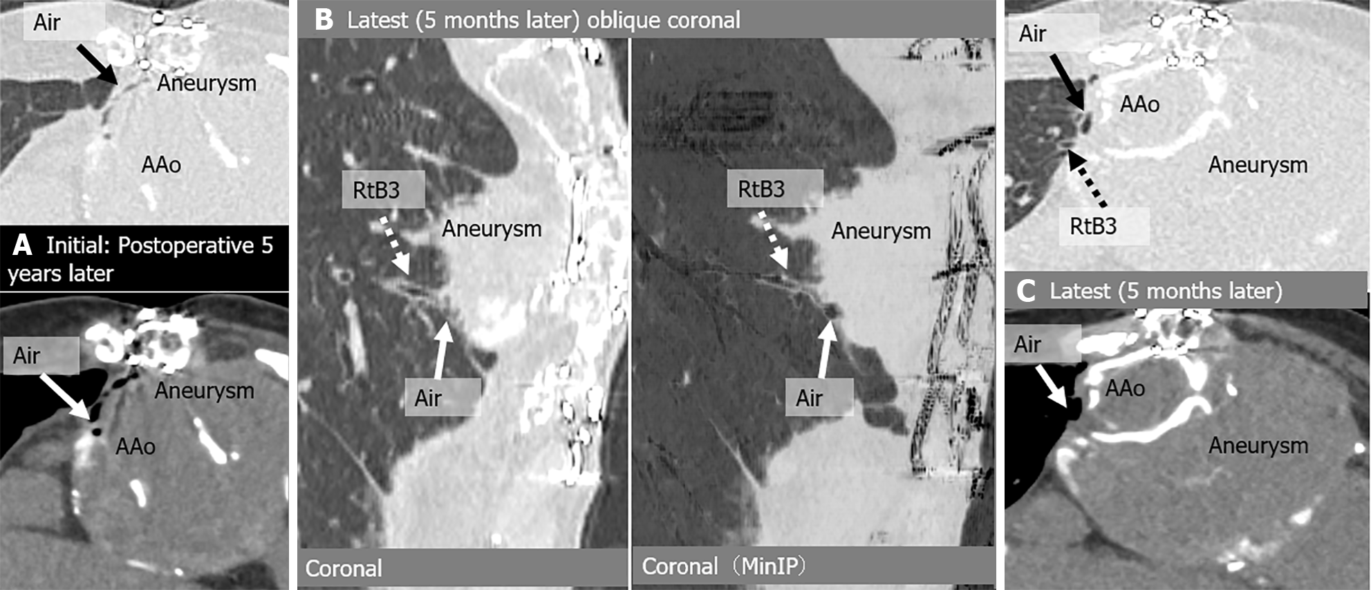
Figure 2 Aortobronchial fistula in an asymptomatic 73-year-old woman with a history of ascending aorta and aortic arch replacement + thoracic endovascular aortic repair 5 years earlier for dissection.
A: Initial computed tomography (CT) images show air shadow in the intra-aortic peri-graft space (arrow); B: Latest CT, 5 months follow-up images show dilated peripheral bronchus (right B3) communicating with peri-graft air(arrow); C: Latest CT, 5-month follow-up images show residual peri-graft air. There were no severe complications during the 5-month observation period. AAo: Ascending aorta; Rt: Right; MinIP: Minimum intensity projection.
- Citation: Tsuchiya N, Inafuku H, Yogi S, Iraha Y, Iida G, Ando M, Nagano T, Higa S, Maeda T, Kise Y, Furukawa K, Yonemoto K, Nishie A. Direct visualization of postoperative aortobronchial fistula on computed tomography. World J Radiol 2024; 16(8): 337-347
- URL: https://www.wjgnet.com/1949-8470/full/v16/i8/337.htm
- DOI: https://dx.doi.org/10.4329/wjr.v16.i8.337


