Copyright
©The Author(s) 2024.
World J Radiol. Aug 28, 2024; 16(8): 337-347
Published online Aug 28, 2024. doi: 10.4329/wjr.v16.i8.337
Published online Aug 28, 2024. doi: 10.4329/wjr.v16.i8.337
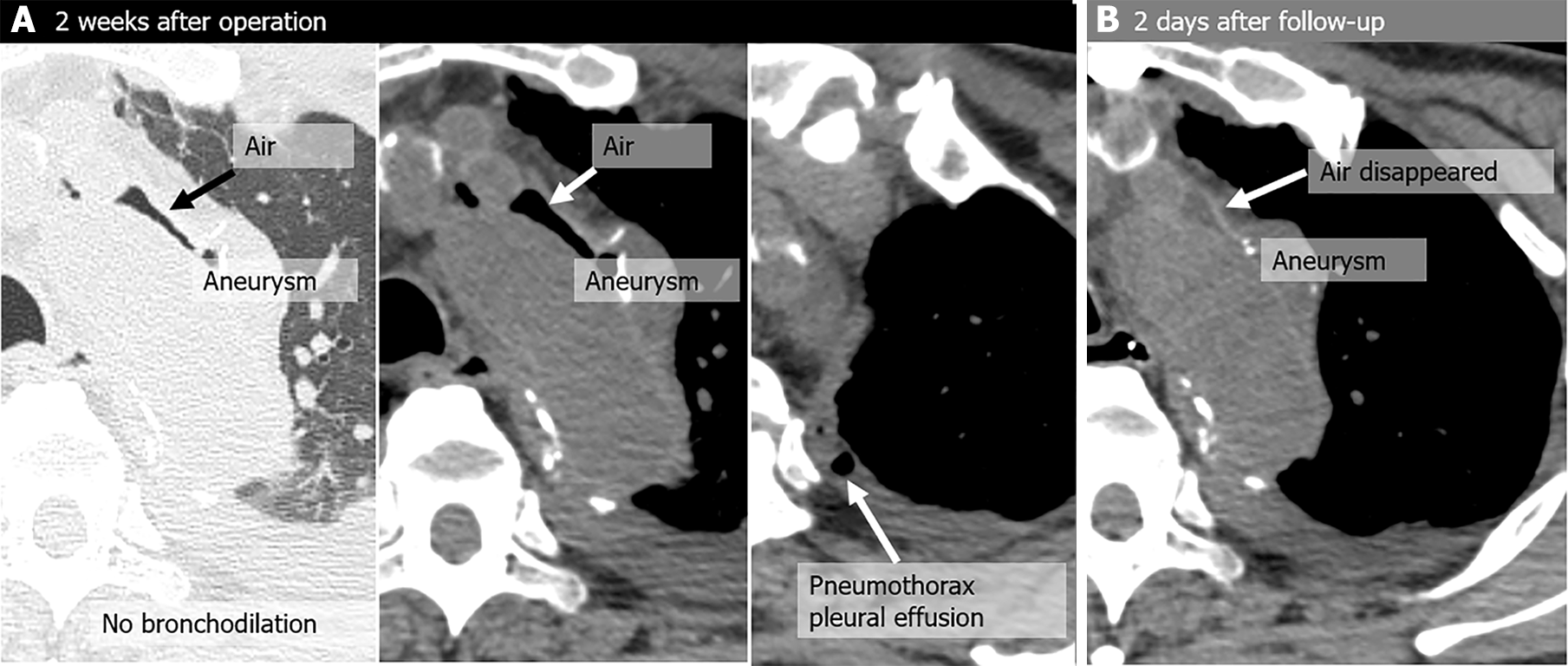
Figure 1 Pseudo-aortobronchial fistula in a 75-year-old man with dyspnea, a history of aortic arch replacement 2 weeks earlier for an aneurysm, and positive pressure ventilation performed for acute respiratory distress syndrome.
A: Two weeks after the operation, he underwent computed tomography (CT) because of dyspnea, and peri-graft air was noted (not seen immediately after the operation); B: The air disappeared on a CT scan performed 2 days later. There was no recurrence of air around the graft on imaging performed over a 6-year period, and no signs of graft infection.
- Citation: Tsuchiya N, Inafuku H, Yogi S, Iraha Y, Iida G, Ando M, Nagano T, Higa S, Maeda T, Kise Y, Furukawa K, Yonemoto K, Nishie A. Direct visualization of postoperative aortobronchial fistula on computed tomography. World J Radiol 2024; 16(8): 337-347
- URL: https://www.wjgnet.com/1949-8470/full/v16/i8/337.htm
- DOI: https://dx.doi.org/10.4329/wjr.v16.i8.337


