Copyright
©The Author(s) 2024.
World J Radiol. Aug 28, 2024; 16(8): 294-316
Published online Aug 28, 2024. doi: 10.4329/wjr.v16.i8.294
Published online Aug 28, 2024. doi: 10.4329/wjr.v16.i8.294
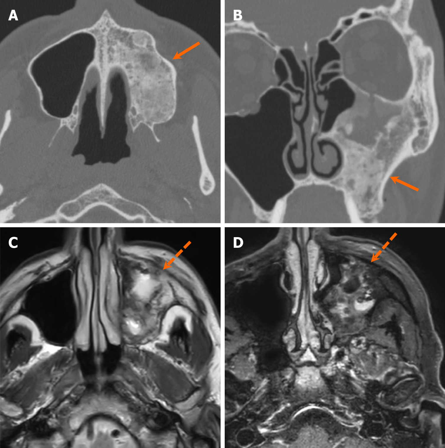
Figure 22 Fibrous dysplasia.
A 62-year-old man was found to have an incidental lesion in the left maxillary sinus wall on magnetic resonance imaging conducted to evaluate sensorineural hearing loss. A and B: Axial (A) and coronal (B) computed tomography images demonstrate an expansile, ground-glass density lesion involving the left maxilla (arrows) which is characteristic of fibrous dysplasia; C and D: Axial T2-weighted (C) and contrast-enhanced fat-suppressed T1-weighted (D) magnetic resonance images show mixed T2 signal intensity and heterogeneous enhancement (dashed arrows). The lesion has remained stable over 7 years of follow-up magnetic resonance imaging.
- Citation: Choi WJ, Lee P, Thomas PC, Rath TJ, Mogensen MA, Dalley RW, Wangaryattawanich P. Imaging approach for jaw and maxillofacial bone tumors with updates from the 2022 World Health Organization classification. World J Radiol 2024; 16(8): 294-316
- URL: https://www.wjgnet.com/1949-8470/full/v16/i8/294.htm
- DOI: https://dx.doi.org/10.4329/wjr.v16.i8.294


