Copyright
©The Author(s) 2024.
World J Radiol. Aug 28, 2024; 16(8): 294-316
Published online Aug 28, 2024. doi: 10.4329/wjr.v16.i8.294
Published online Aug 28, 2024. doi: 10.4329/wjr.v16.i8.294
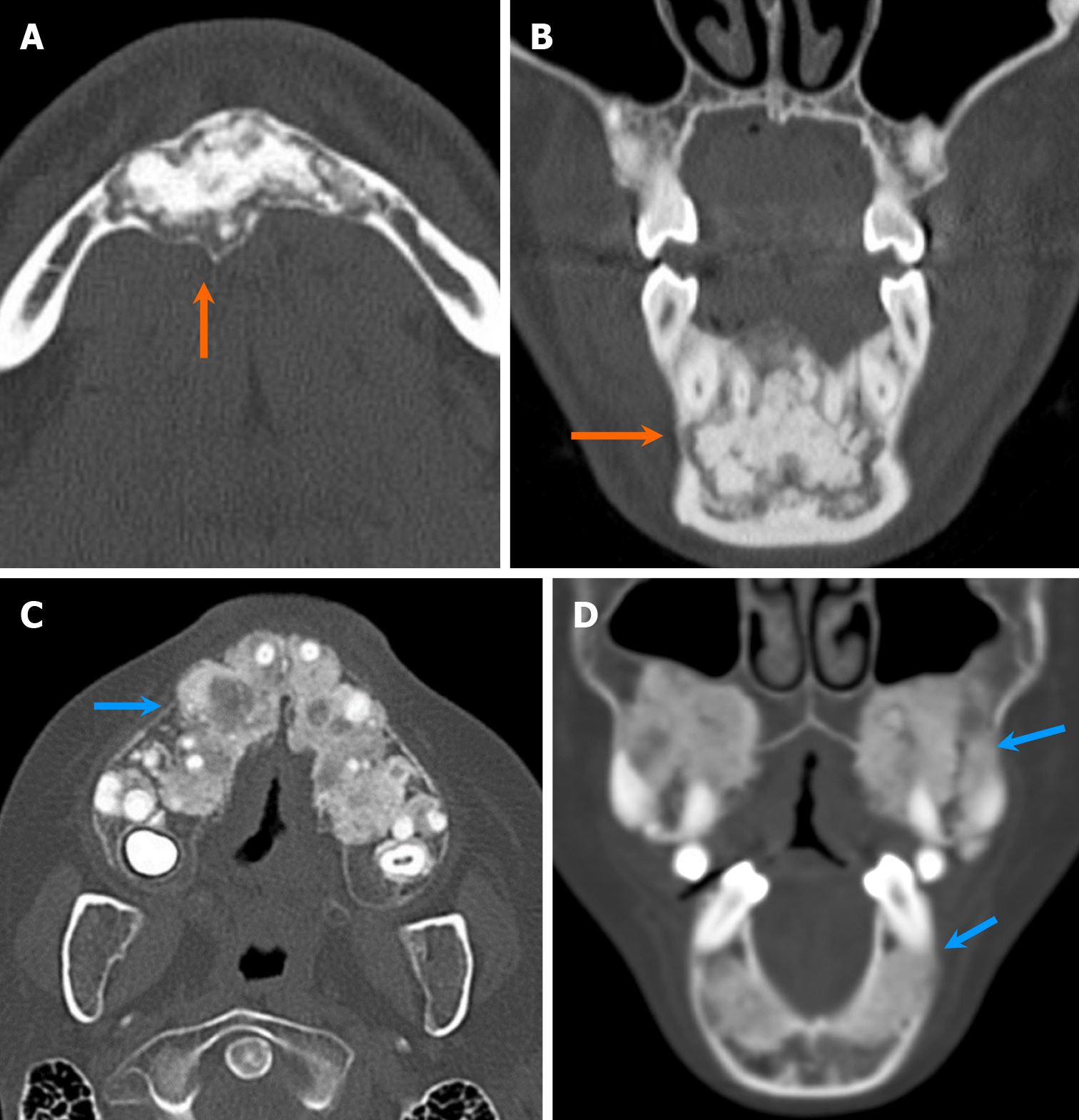
Figure 21 Two cases of cemento-osseous dysplasia.
A and B: Axial (A) and coronal (B) computed tomography images show a solitary, mildly expansile, densely sclerotic lesion in the anterior mandible with a narrow radiolucent rim and well-circumscribed borders (orange arrows). The lesions are attached to the roots of multiple teeth, indicating periapical cemento-osseous dysplasia; C and D: Axial (C) and coronal (D) computed tomography images of a companion case depict multifocal lesions with a ground-glass matrix in both maxilla and mandible (blue arrows), indicative of florid cemento-osseous dysplasia.
- Citation: Choi WJ, Lee P, Thomas PC, Rath TJ, Mogensen MA, Dalley RW, Wangaryattawanich P. Imaging approach for jaw and maxillofacial bone tumors with updates from the 2022 World Health Organization classification. World J Radiol 2024; 16(8): 294-316
- URL: https://www.wjgnet.com/1949-8470/full/v16/i8/294.htm
- DOI: https://dx.doi.org/10.4329/wjr.v16.i8.294


