Copyright
©The Author(s) 2024.
World J Radiol. Aug 28, 2024; 16(8): 294-316
Published online Aug 28, 2024. doi: 10.4329/wjr.v16.i8.294
Published online Aug 28, 2024. doi: 10.4329/wjr.v16.i8.294
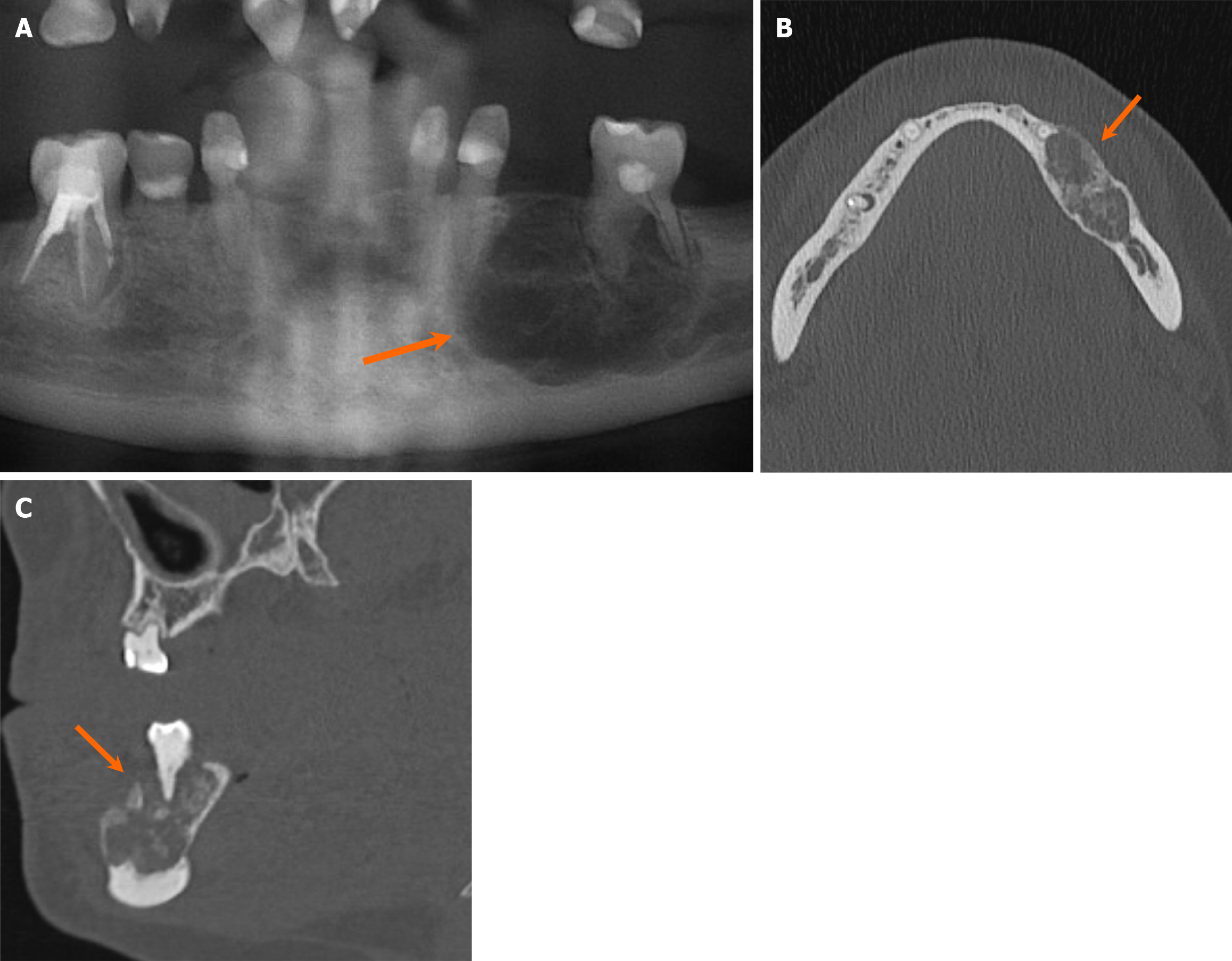
Figure 20 Cemento-ossifying fibroma.
A 34-year-old woman with an incidentally discovered radiolucent mandibular lesion. A-C: Orthopantomogram (A), axial (B) and sagittal (C) computed tomography images demonstrate a mixed lucent and ground-glass, mildly expansile lesion with well-circumscribed borders, involving the left mandibular body (arrows). Pathology confirmed cemento-ossifying fibroma following resection.
- Citation: Choi WJ, Lee P, Thomas PC, Rath TJ, Mogensen MA, Dalley RW, Wangaryattawanich P. Imaging approach for jaw and maxillofacial bone tumors with updates from the 2022 World Health Organization classification. World J Radiol 2024; 16(8): 294-316
- URL: https://www.wjgnet.com/1949-8470/full/v16/i8/294.htm
- DOI: https://dx.doi.org/10.4329/wjr.v16.i8.294


