Copyright
©The Author(s) 2024.
World J Radiol. Aug 28, 2024; 16(8): 294-316
Published online Aug 28, 2024. doi: 10.4329/wjr.v16.i8.294
Published online Aug 28, 2024. doi: 10.4329/wjr.v16.i8.294
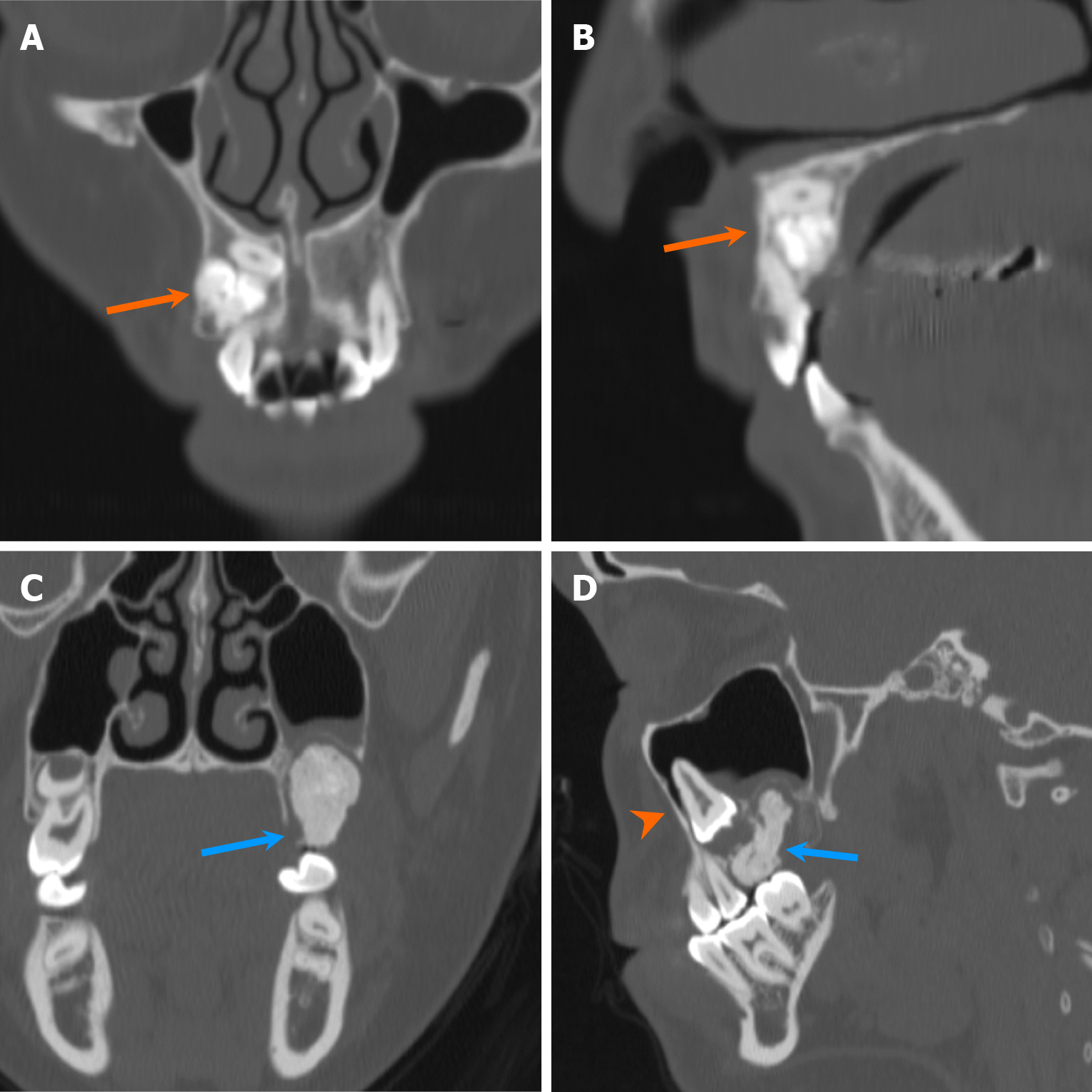
Figure 18 Compound and complex odontomas.
A and B: Coronal (A) and sagittal (B) computed tomography images show a densely sclerotic lesion in the right maxilla (arrows), composed of several small denticles resembling a tooth, consistent with a compound odontoma; C and D: Coronal (C) and sagittal (D) computed tomography images reveal an amorphous, densely sclerotic lesion in the left maxilla with a lucent rim (blue arrows), consistent with a complex odontoma. Note the displaced unerupted tooth just above the lesion (arrowhead).
- Citation: Choi WJ, Lee P, Thomas PC, Rath TJ, Mogensen MA, Dalley RW, Wangaryattawanich P. Imaging approach for jaw and maxillofacial bone tumors with updates from the 2022 World Health Organization classification. World J Radiol 2024; 16(8): 294-316
- URL: https://www.wjgnet.com/1949-8470/full/v16/i8/294.htm
- DOI: https://dx.doi.org/10.4329/wjr.v16.i8.294


