Copyright
©The Author(s) 2024.
World J Radiol. Aug 28, 2024; 16(8): 294-316
Published online Aug 28, 2024. doi: 10.4329/wjr.v16.i8.294
Published online Aug 28, 2024. doi: 10.4329/wjr.v16.i8.294
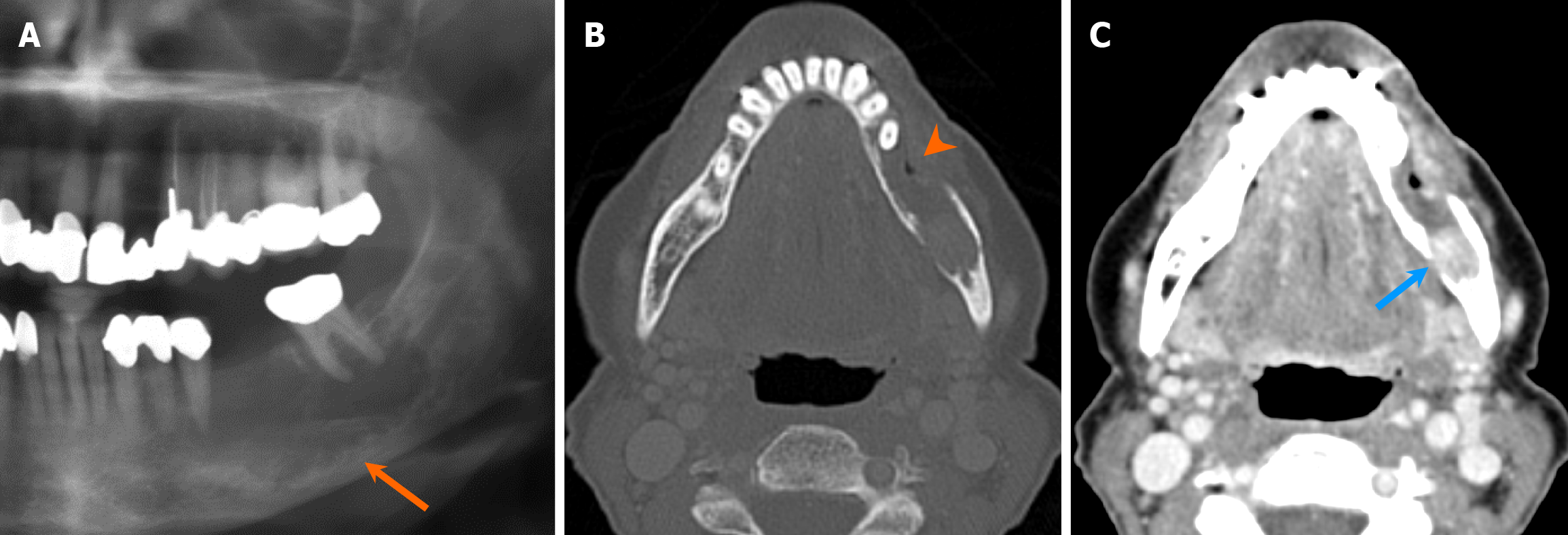
Figure 16 Odontogenic carcinoma.
A 71-year-old woman with persistent dull aching pain of the left mandible and left V3 paresthesia. A-C: Orthopantomogram (A) reveals a radiolucent lesion with ill-defined margins in the mandibular body, involving the inferior alveolar canal (arrow). Axial computed tomography images in bone (B) and soft tissue (C) windows show extensive cortical destruction (arrowhead) and an enhancing soft tissue component within the lesion (blue arrow). Pathology confirmed odontogenic carcinoma following resection.
- Citation: Choi WJ, Lee P, Thomas PC, Rath TJ, Mogensen MA, Dalley RW, Wangaryattawanich P. Imaging approach for jaw and maxillofacial bone tumors with updates from the 2022 World Health Organization classification. World J Radiol 2024; 16(8): 294-316
- URL: https://www.wjgnet.com/1949-8470/full/v16/i8/294.htm
- DOI: https://dx.doi.org/10.4329/wjr.v16.i8.294


