Copyright
©The Author(s) 2024.
World J Radiol. Aug 28, 2024; 16(8): 294-316
Published online Aug 28, 2024. doi: 10.4329/wjr.v16.i8.294
Published online Aug 28, 2024. doi: 10.4329/wjr.v16.i8.294
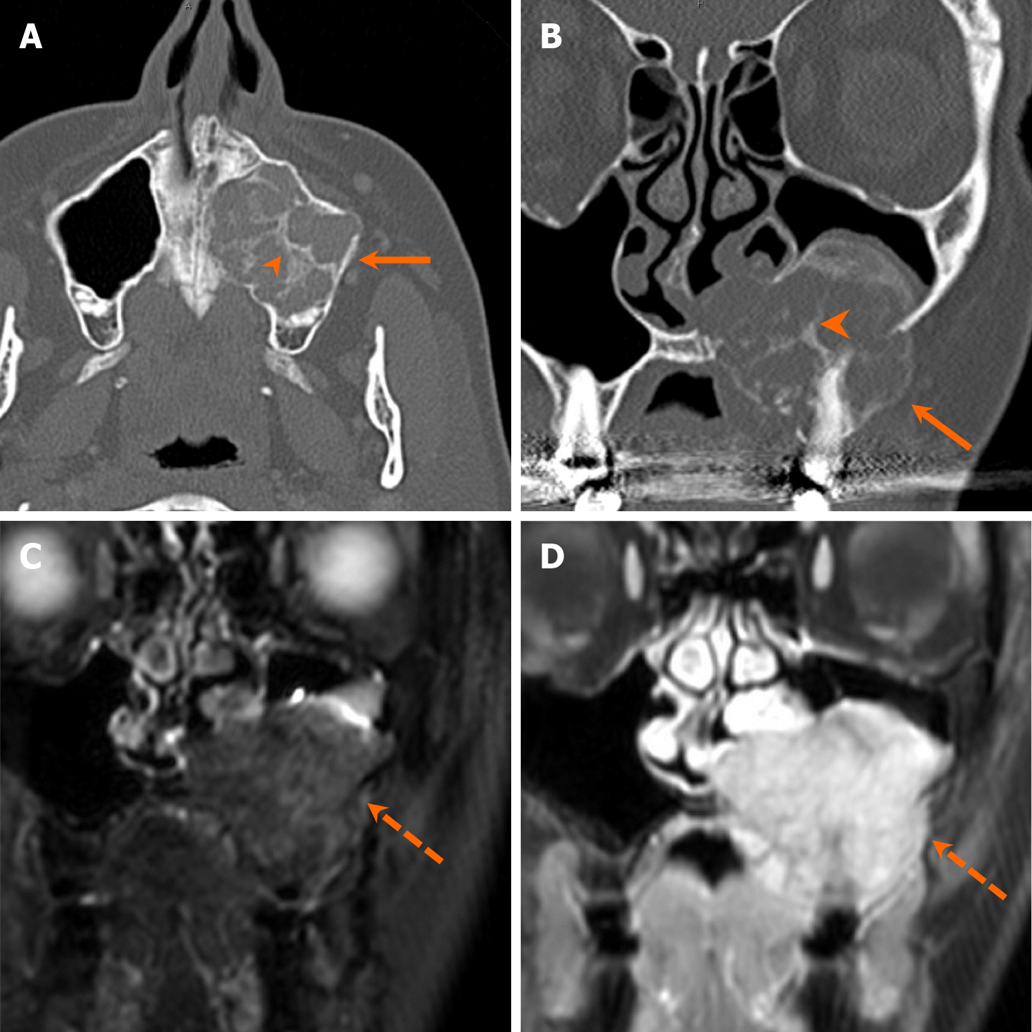
Figure 15 Giant cell granuloma.
A 58-year-old woman with left cheek discomfort. A and B: Axial (A) and coronal (B) computed tomography images show an expansile, multilocular radiolucent lesion involving the maxillary alveolus (arrows), with visible irregular ground-glass septa (arrowheads); C and D: Coronal STIR (C) and contrast-enhanced fat-suppressed T1-weighted (D) magnetic resonance images reveal an avidly enhancing, T2 hyperintense mass (dashed arrows). Pathology confirmed giant cell granuloma following resection.
- Citation: Choi WJ, Lee P, Thomas PC, Rath TJ, Mogensen MA, Dalley RW, Wangaryattawanich P. Imaging approach for jaw and maxillofacial bone tumors with updates from the 2022 World Health Organization classification. World J Radiol 2024; 16(8): 294-316
- URL: https://www.wjgnet.com/1949-8470/full/v16/i8/294.htm
- DOI: https://dx.doi.org/10.4329/wjr.v16.i8.294


