Copyright
©The Author(s) 2024.
World J Radiol. Aug 28, 2024; 16(8): 294-316
Published online Aug 28, 2024. doi: 10.4329/wjr.v16.i8.294
Published online Aug 28, 2024. doi: 10.4329/wjr.v16.i8.294
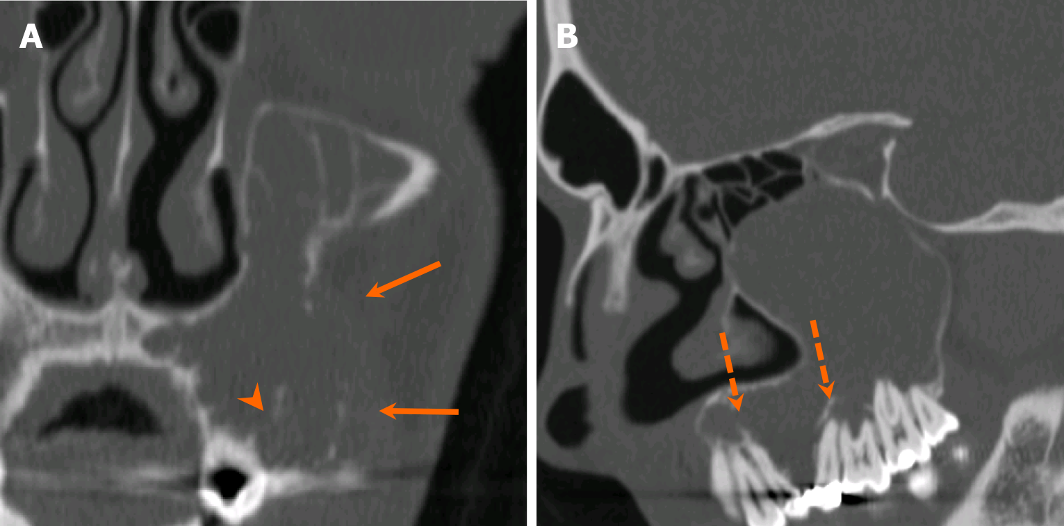
Figure 14 Odontogenic myxoma.
A 27-year-old man with a slowly enlarging, nonpainful left maxillary alveolar mass for several years. A and B: Coronal (A) and sagittal (B) computed tomography images reveal a large, expansile, multilocular radiolucent lesion originating from the maxillary alveolus and extending into the maxillary sinus, causing bone destruction with multifocal dehiscence (arrows). Note the ill-defined margins with internal septations along the alveolar process (arrowhead). The mass exerts a mass effect, causing tilting of the adjacent teeth (dashed arrows). Pathology confirmed odontogenic myxoma following resection.
- Citation: Choi WJ, Lee P, Thomas PC, Rath TJ, Mogensen MA, Dalley RW, Wangaryattawanich P. Imaging approach for jaw and maxillofacial bone tumors with updates from the 2022 World Health Organization classification. World J Radiol 2024; 16(8): 294-316
- URL: https://www.wjgnet.com/1949-8470/full/v16/i8/294.htm
- DOI: https://dx.doi.org/10.4329/wjr.v16.i8.294


