Copyright
©The Author(s) 2024.
World J Radiol. Aug 28, 2024; 16(8): 294-316
Published online Aug 28, 2024. doi: 10.4329/wjr.v16.i8.294
Published online Aug 28, 2024. doi: 10.4329/wjr.v16.i8.294
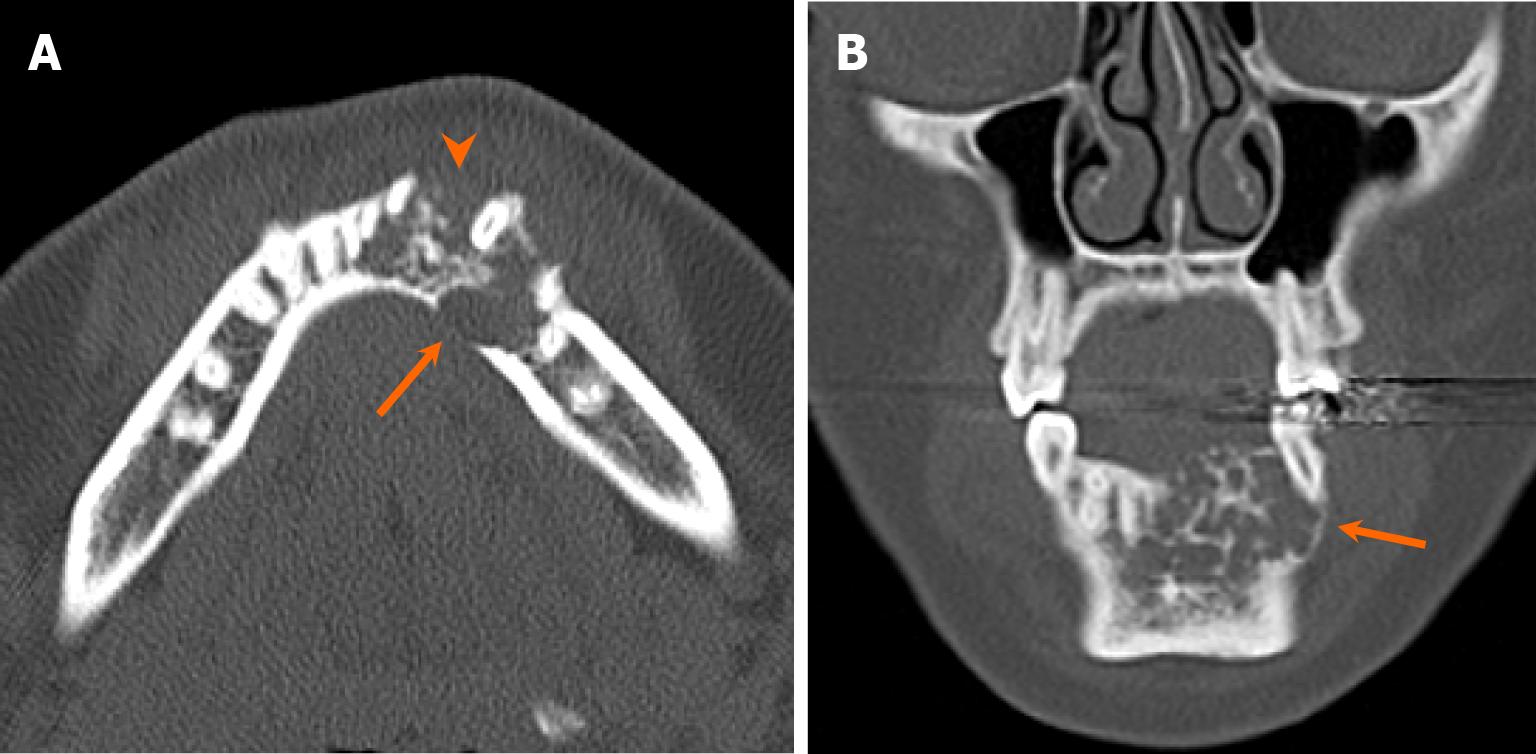
Figure 12 Conventional ameloblastoma of the mandible.
A 17-year-old girl with a several-month history of mandibular swelling and bilateral facial pain. A and B: Axial (A) and coronal (B) computed tomography images reveal an expansile multilocular radiolucent lesion in the left anterior aspect of the mandible (arrows), with a soap-bubble appearance. Note the multifocal cortical dehiscence in the affected bone (arrowhead). Patient underwent surgical resection with free flap reconstruction, and final pathology confirmed ameloblastoma.
- Citation: Choi WJ, Lee P, Thomas PC, Rath TJ, Mogensen MA, Dalley RW, Wangaryattawanich P. Imaging approach for jaw and maxillofacial bone tumors with updates from the 2022 World Health Organization classification. World J Radiol 2024; 16(8): 294-316
- URL: https://www.wjgnet.com/1949-8470/full/v16/i8/294.htm
- DOI: https://dx.doi.org/10.4329/wjr.v16.i8.294


