Copyright
©The Author(s) 2024.
World J Radiol. Aug 28, 2024; 16(8): 294-316
Published online Aug 28, 2024. doi: 10.4329/wjr.v16.i8.294
Published online Aug 28, 2024. doi: 10.4329/wjr.v16.i8.294
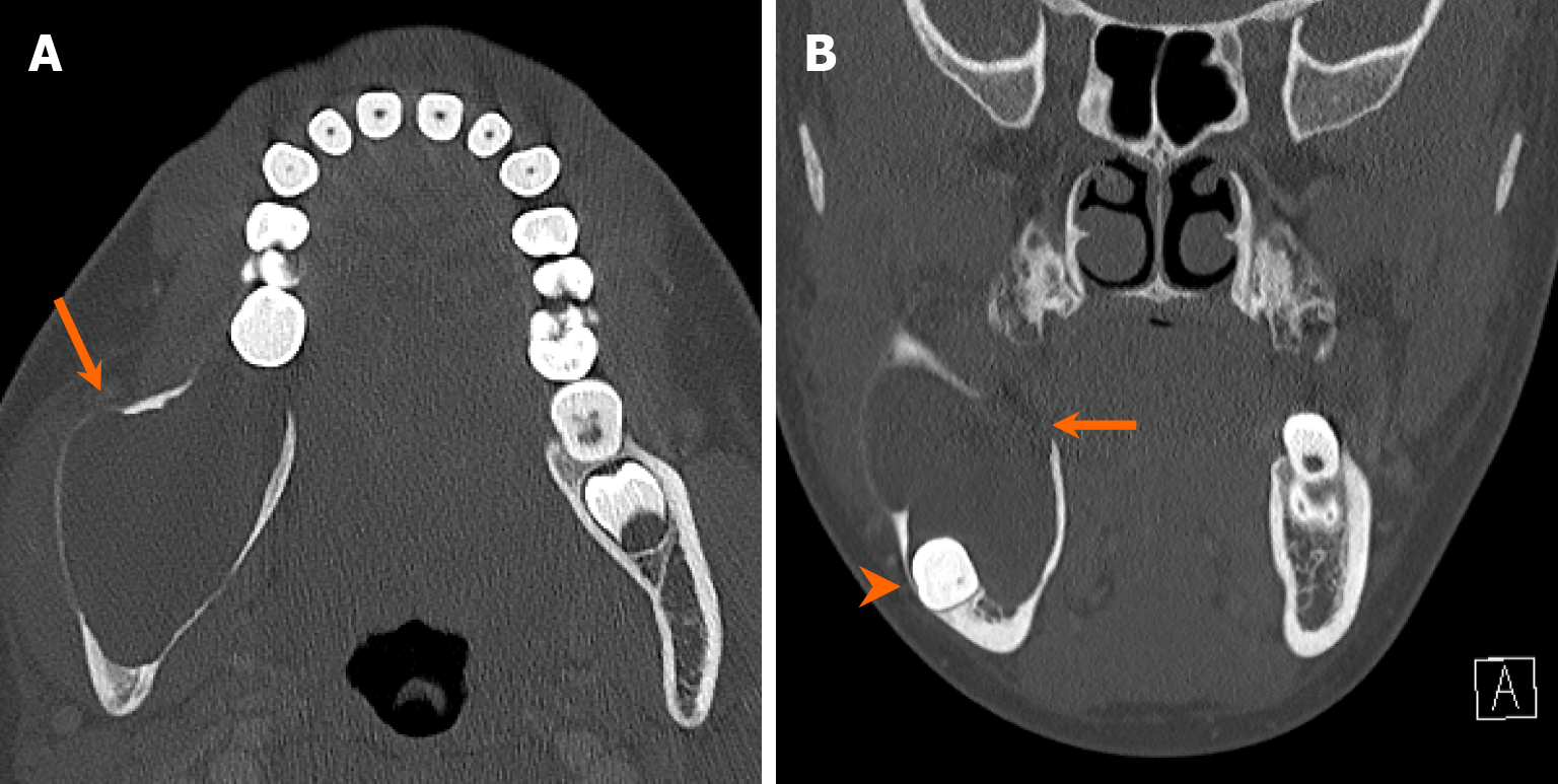
Figure 11 Unicystic ameloblastoma.
An 11-year-old girl with a 2-week history of right lower jaw swelling and facial pain. A and B: Axial (A) and coronal (B) computed tomography images demonstrate a large, expansile, unilocular, cystic lesion centered at the right mandibular angle (arrows). There is associated cortical thinning and multifocal bone dehiscence. Notably, there is inferior displacement of the unerupted third molar due to mass effect (arrowhead). Patient underwent surgical resection, with a pathologically confirmed diagnosis of unicystic ameloblastoma.
- Citation: Choi WJ, Lee P, Thomas PC, Rath TJ, Mogensen MA, Dalley RW, Wangaryattawanich P. Imaging approach for jaw and maxillofacial bone tumors with updates from the 2022 World Health Organization classification. World J Radiol 2024; 16(8): 294-316
- URL: https://www.wjgnet.com/1949-8470/full/v16/i8/294.htm
- DOI: https://dx.doi.org/10.4329/wjr.v16.i8.294


