Copyright
©The Author(s) 2024.
World J Radiol. Aug 28, 2024; 16(8): 294-316
Published online Aug 28, 2024. doi: 10.4329/wjr.v16.i8.294
Published online Aug 28, 2024. doi: 10.4329/wjr.v16.i8.294
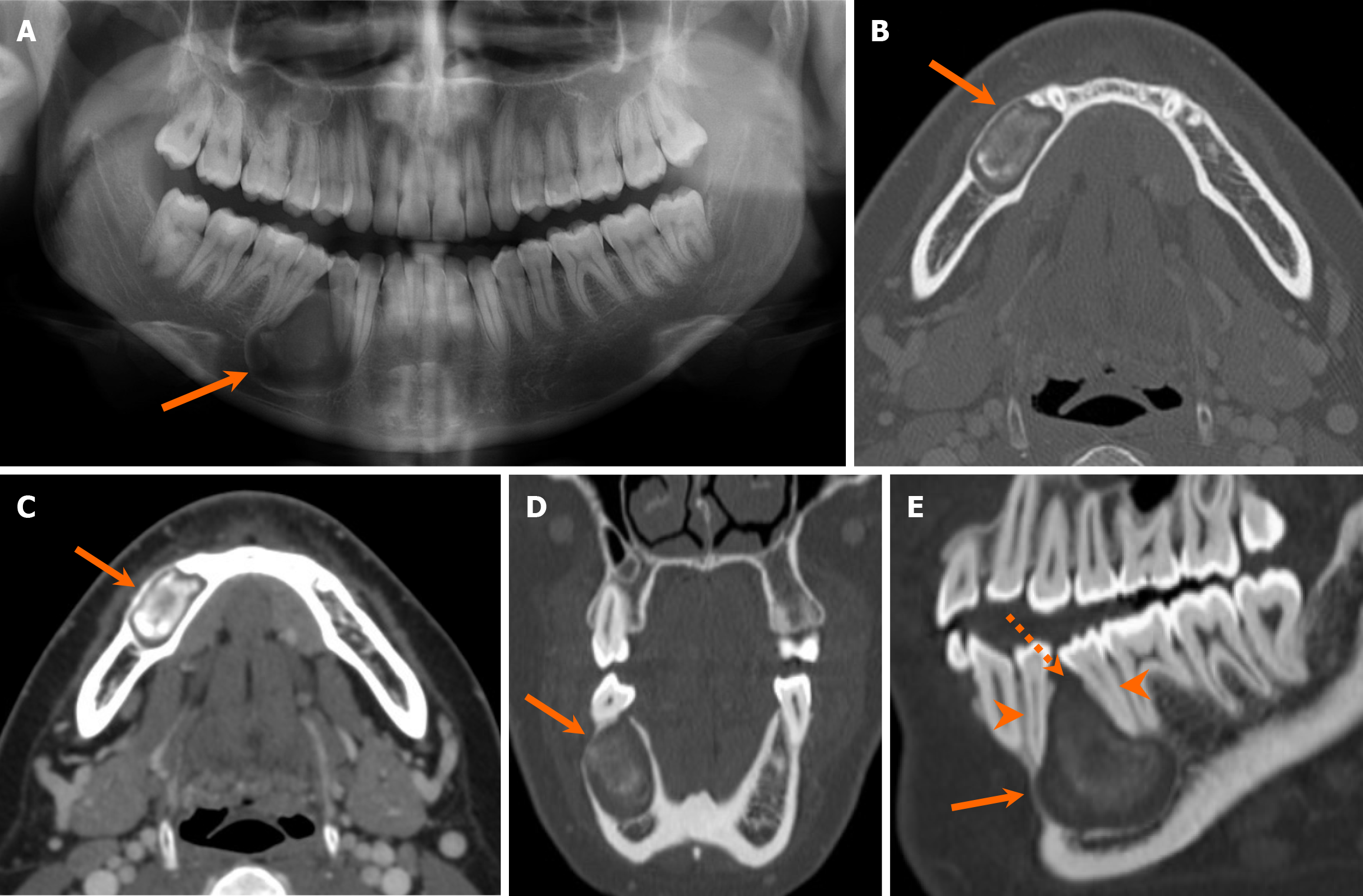
Figure 10 Odontogenic keratocyst.
A 23-year-old woman with an incidental radiolucent mandibular lesion detected on an X-ray. A-E: The orthopantomogram (A), axial (B and C), coronal (D), and sagittal oblique (E) computed tomography images demonstrate an expansile unilocular radiolucent lesion centered in the right mandibular body (arrows), containing mixed-density content. The lesion extends along the longitudinal axis of the mandible and exerts mass effect, causing mild displacement but no erosion of the adjacent teeth (arrowheads). There is focal cortical dehiscence at the superior aspect of the lesion (dashed arrow). Patient underwent surgical resection, and final pathology confirmed an odontogenic keratocyst.
- Citation: Choi WJ, Lee P, Thomas PC, Rath TJ, Mogensen MA, Dalley RW, Wangaryattawanich P. Imaging approach for jaw and maxillofacial bone tumors with updates from the 2022 World Health Organization classification. World J Radiol 2024; 16(8): 294-316
- URL: https://www.wjgnet.com/1949-8470/full/v16/i8/294.htm
- DOI: https://dx.doi.org/10.4329/wjr.v16.i8.294


