Copyright
©The Author(s) 2024.
World J Radiol. Aug 28, 2024; 16(8): 294-316
Published online Aug 28, 2024. doi: 10.4329/wjr.v16.i8.294
Published online Aug 28, 2024. doi: 10.4329/wjr.v16.i8.294
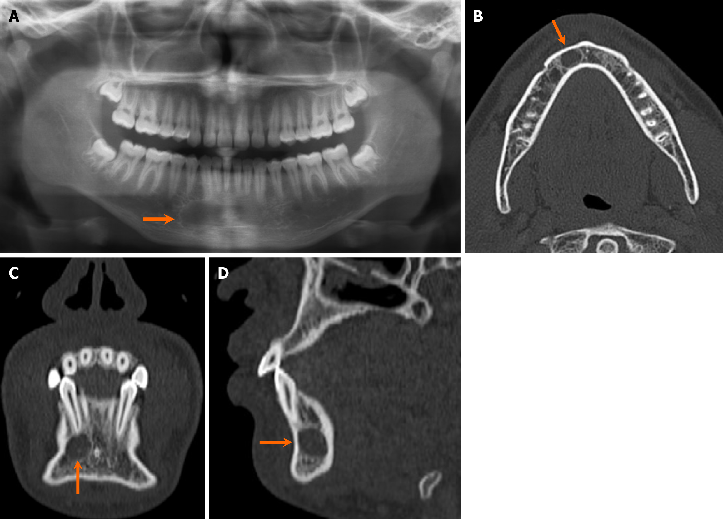
Figure 9 Simple bone cyst (aka traumatic bone cyst).
An 18-year-old man with an incidentally discovered radiolucent lesion in the right mandible. He has reported no associated pain, bony expansion, drainage, or numbness. A-D: Orthopantomogram (A), axial (B), coronal (C), and sagittal (D) computed tomography images reveal a small, well-marginated, unilocular cystic lesion at the right parasymphysis of the mandible, situated inferior to the apex of the lateral incisor (arrows), with no evidence of bone expansion. The tooth appears intact, showing no signs of erosion or associated dental caries. Patient underwent exploration and curettage of the lesion, with pathological confirmation of a simple bone cyst.
- Citation: Choi WJ, Lee P, Thomas PC, Rath TJ, Mogensen MA, Dalley RW, Wangaryattawanich P. Imaging approach for jaw and maxillofacial bone tumors with updates from the 2022 World Health Organization classification. World J Radiol 2024; 16(8): 294-316
- URL: https://www.wjgnet.com/1949-8470/full/v16/i8/294.htm
- DOI: https://dx.doi.org/10.4329/wjr.v16.i8.294


