Copyright
©The Author(s) 2024.
World J Radiol. Aug 28, 2024; 16(8): 294-316
Published online Aug 28, 2024. doi: 10.4329/wjr.v16.i8.294
Published online Aug 28, 2024. doi: 10.4329/wjr.v16.i8.294
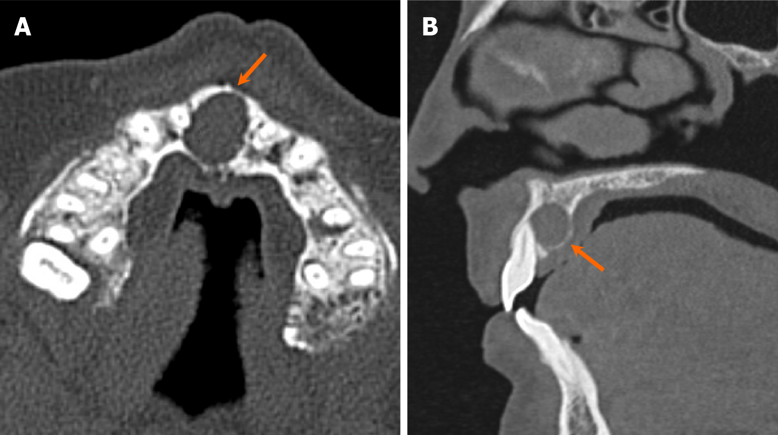
Figure 8 Nasopalatine duct cyst.
A and B: Axial (A) and sagittal (B) computed tomography images demonstrate an expansile, unilocular, well-marginated, cystic lesion located in the region of the incisive foramen (arrows). These findings are highly indicative of a nasopalatine duct cyst, a developmental non-odontogenic cyst resulting from incomplete regression of epithelium in the nasopalatine duct.
- Citation: Choi WJ, Lee P, Thomas PC, Rath TJ, Mogensen MA, Dalley RW, Wangaryattawanich P. Imaging approach for jaw and maxillofacial bone tumors with updates from the 2022 World Health Organization classification. World J Radiol 2024; 16(8): 294-316
- URL: https://www.wjgnet.com/1949-8470/full/v16/i8/294.htm
- DOI: https://dx.doi.org/10.4329/wjr.v16.i8.294


