Copyright
©The Author(s) 2024.
World J Radiol. Aug 28, 2024; 16(8): 294-316
Published online Aug 28, 2024. doi: 10.4329/wjr.v16.i8.294
Published online Aug 28, 2024. doi: 10.4329/wjr.v16.i8.294
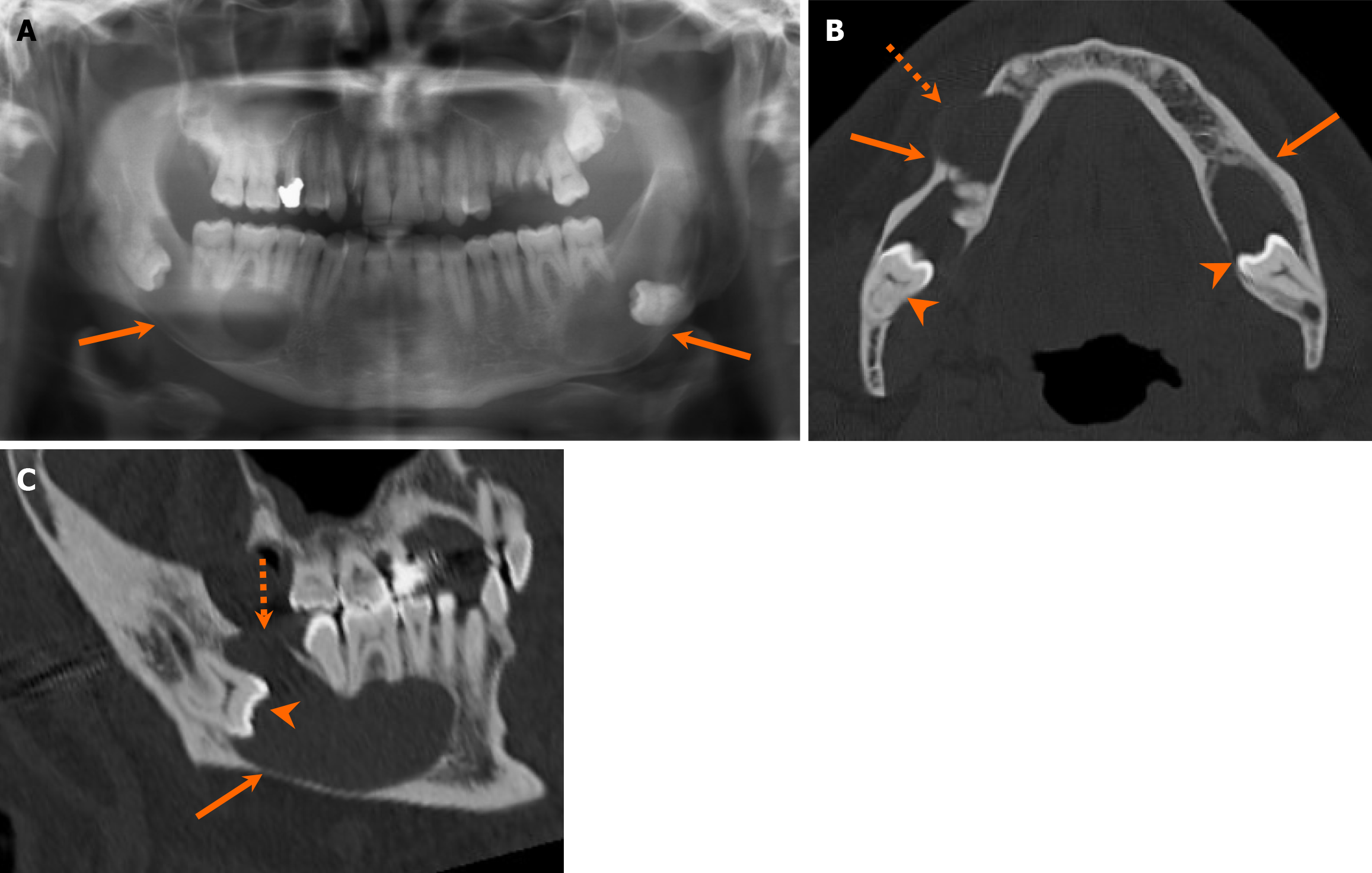
Figure 7 Dentigerous cyst.
A 51-year-old man with 6-month history of jaw swelling and liquid intermittently draining into his mouth. A-C: Orthopantomogram (A), axial (B), and sagittal (C) computed tomography images reveal large unilocular cystic lesions in the bilateral mandibular bodies (arrows) centered at the crown of unerupted molar teeth (arrowheads), characteristic features of the dentigerous cyst. The lesions expand along the longitudinal axis (i.e., anteroposterior dimension) of the mandible, with focal bone dehiscence in multiple areas (dashed arrows). Patient underwent surgical resection, with pathologically confirmed dentigerous cysts.
- Citation: Choi WJ, Lee P, Thomas PC, Rath TJ, Mogensen MA, Dalley RW, Wangaryattawanich P. Imaging approach for jaw and maxillofacial bone tumors with updates from the 2022 World Health Organization classification. World J Radiol 2024; 16(8): 294-316
- URL: https://www.wjgnet.com/1949-8470/full/v16/i8/294.htm
- DOI: https://dx.doi.org/10.4329/wjr.v16.i8.294


