Copyright
©The Author(s) 2024.
World J Radiol. Aug 28, 2024; 16(8): 294-316
Published online Aug 28, 2024. doi: 10.4329/wjr.v16.i8.294
Published online Aug 28, 2024. doi: 10.4329/wjr.v16.i8.294
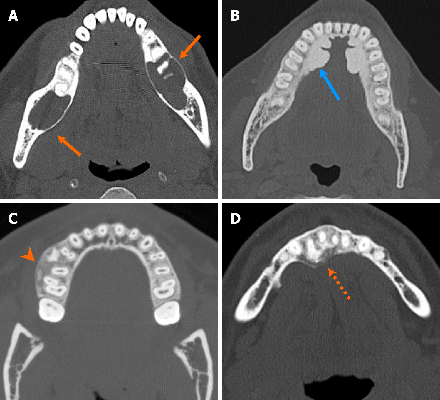
Figure 4 Radiodensity types of the jaw and maxillofacial bone lesions.
Axial computed tomography images of four cases illustrate various types of radiodensity of the jaw lesions. A: Radiolucent lesion in a patient with multiple simple bone cysts (orange arrows); B: Densely sclerotic lesion in a patient with torus mandibularis (blue arrow); C: Ground-glass density lesion in a patient with psammomatoid ossifying fibroma (arrowhead); D: Mixed lytic and sclerotic lesion in a patient with cemento-osseous dysplasia (dash arrow).
- Citation: Choi WJ, Lee P, Thomas PC, Rath TJ, Mogensen MA, Dalley RW, Wangaryattawanich P. Imaging approach for jaw and maxillofacial bone tumors with updates from the 2022 World Health Organization classification. World J Radiol 2024; 16(8): 294-316
- URL: https://www.wjgnet.com/1949-8470/full/v16/i8/294.htm
- DOI: https://dx.doi.org/10.4329/wjr.v16.i8.294


