Copyright
©The Author(s) 2024.
World J Radiol. Aug 28, 2024; 16(8): 294-316
Published online Aug 28, 2024. doi: 10.4329/wjr.v16.i8.294
Published online Aug 28, 2024. doi: 10.4329/wjr.v16.i8.294
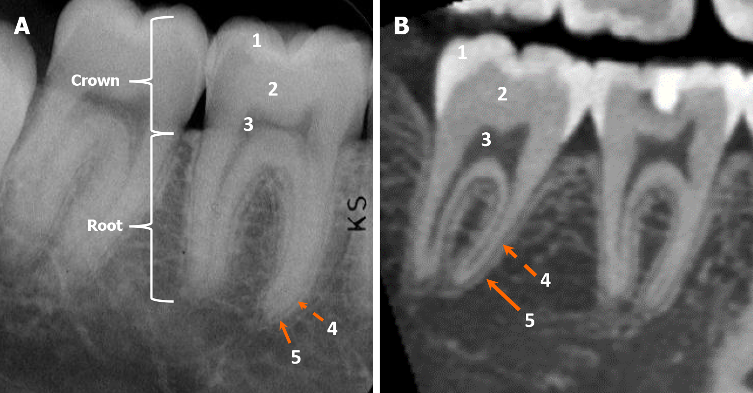
Figure 3 Dental anatomy.
A and B: The intraoral radiograph (A) and cone beam computed tomography parasagittal view (B) demonstrate normal dental anatomy with distinct anatomical features, including: (1) Enamel; (2) Dentin; (3) Pulp cavity; (4) Periodontal ligament (radiolucent line); and (5) Lamina dura (radiodense line). Radiologically, cementum appears isodense to dentin; therefore, cementum and dentin cannot be distinguished on radiographs or computed tomography.
- Citation: Choi WJ, Lee P, Thomas PC, Rath TJ, Mogensen MA, Dalley RW, Wangaryattawanich P. Imaging approach for jaw and maxillofacial bone tumors with updates from the 2022 World Health Organization classification. World J Radiol 2024; 16(8): 294-316
- URL: https://www.wjgnet.com/1949-8470/full/v16/i8/294.htm
- DOI: https://dx.doi.org/10.4329/wjr.v16.i8.294


