Copyright
©The Author(s) 2024.
World J Radiol. Aug 28, 2024; 16(8): 294-316
Published online Aug 28, 2024. doi: 10.4329/wjr.v16.i8.294
Published online Aug 28, 2024. doi: 10.4329/wjr.v16.i8.294
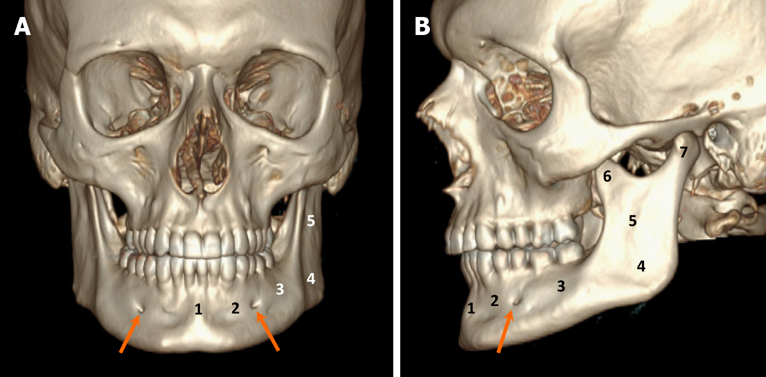
Figure 1 Anatomy of the mandible.
A and B: Computed tomography images with volume rendering reconstruction in anteroposterior (A) and lateral (B) views highlighting distinct mandibular regions: (1) Symphysis; (2) Parasymphysis; (3) Body; (4) Angle; (5) Ramus; (6) Coronoid process; and (7) Condylar process. Note the mental foramen (arrows), serving as exit points for the inferior alveolar nerves from the mandible, situated proximate to the first and second premolar teeth.
- Citation: Choi WJ, Lee P, Thomas PC, Rath TJ, Mogensen MA, Dalley RW, Wangaryattawanich P. Imaging approach for jaw and maxillofacial bone tumors with updates from the 2022 World Health Organization classification. World J Radiol 2024; 16(8): 294-316
- URL: https://www.wjgnet.com/1949-8470/full/v16/i8/294.htm
- DOI: https://dx.doi.org/10.4329/wjr.v16.i8.294


